. 2014 Nov 19;34(47):15601-9.
doi: 10.1523/JNEUROSCI.2664-14.2014.

Pathway-selective adjustment of prefrontal-amygdala transmission during fear encoding

Affiliations

Pathway-selective adjustment of prefrontal-amygdala transmission during fear encoding

Maithe Arruda-Carvalho et al. J Neurosci. .

Abstract

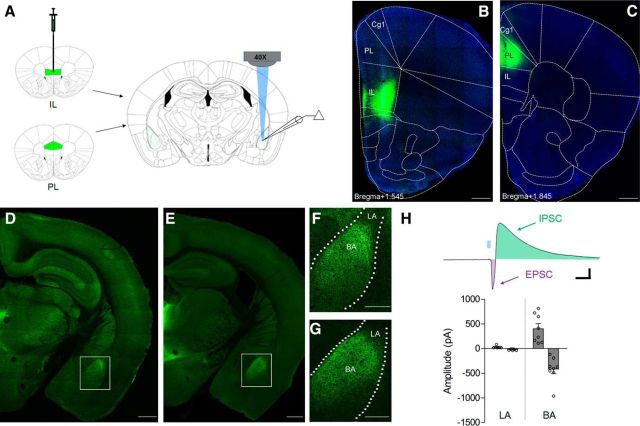
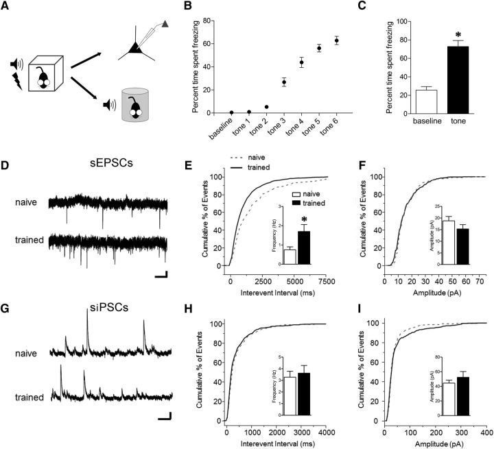
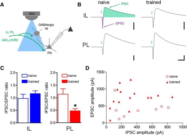
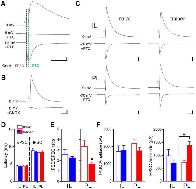
Conditioned fear requires neural activity in the basolateral amygdala (BLA) and medial prefrontal cortex (mPFC), structures that are densely interconnected at the synaptic level. Previous work has suggested that anatomical subdivisions of mPFC make distinct contributions to fear expression and inhibition, and that the functional output of this processing is relayed to the BLA complex. However, it remains unknown whether synaptic plasticity in mPFC-BLA networks contributes to fear memory encoding. Here we use optogenetics and ex vivo electrophysiology to reveal the impact of fear conditioning on BLA excitatory and feedforward inhibitory circuits formed by projections from infralimbic (IL) and prelimbic (PL) cortices. In naive mice, these pathways recruit equivalent excitation and feedforward inhibition in BLA principal neurons. However, fear learning leads to a selective decrease in inhibition:excitation balance in PL circuits that is attributable to a postsynaptic increase in AMPA receptor function. These data suggest a pathway-specific mechanism for fear memory encoding by adjustment of mPFC-BLA transmission. Upon reengagement of PL by conditioned cues, these modifications may serve to amplify emotional responses.

Keywords: amygdala; channelrhodopsin; fear; memory; prefrontal; synaptic plasticity.


Figures

Figure 1.
Figure 1.
Glutamatergic projections from mPFC recruit excitatory and inhibitory transmission predominantly in the basal amygdala. A, Stereotaxic injections of AAV2-CaMKIIα-ChR2(H134R)-EYFP were targeted to the IL or PL subregions of mPFC 6 weeks before histology or terminal-specific optogenetic electrophysiology in the BLA. B, C, Representative examples of EYFP immunofluorescence in IL (B) or PL targets (C) with DAPI overlay. D–G, Confocal images of terminal-associated EYFP fluorescence in midline thalamic nuclei and BLA after IL (D, F) or PL injection (E, G). Insets depict enrichment of terminals in the basal (BA), but not the lateral (LA) nucleus of the amygdala (F, G). Scale bars: D, E, 500 μm; F, G, 250 μm. H, Optic stimulation of PL terminals (5 ms, 460 nm) evoked large-amplitude biphasic excitatory-inhibitory synaptic responses in principal neurons of the basal, but not the lateral nucleus. Calibration: 60 pA × 40 ms.
Figure 2.
Figure 2.
Limited impact of fear conditioning on spontaneous neurotransmission in the basal nucleus. A, Schedule for obtaining electrophysiological or behavioral measurements for all trained mice. Training entailed six pairings of pure-tone CS (2 kHz, 20 s, 80 dB) with scrambled footshock US (0.7 mA, 2 s), while naive mice were selected from cage-experienced littermates. B, C, Freezing during fear conditioning (B) and retrieval test (C). *p < 0.0001, paired t test versus baseline (n = 10). D, Representative sEPSC traces at −60 mV. Calibration: 10 pA × 400 ms. E, F, Cumulative distribution and group means for frequency (E) and amplitude (F) of sEPSCs, *p < 0.05, two-tailed t test [n = 12 (5) naive, 14 (4) trained]. G, Representative sIPSC traces at 0 mV. Calibration: 30 pA × 300 ms. H, I, Cumulative distribution and group means for frequency (H) and amplitude (I) of sIPSCs [n = 9 (3) naive, 11 (3) trained].
Figure 3.
Figure 3.
Fear conditioning reduces the balance of inhibition to excitation in the PL-amygdala pathway. A, Hypothetical model of glutamatergic excitation and local feedforward inhibition in principal neurons resulting from optic stimulation of IL or PL BLA terminals onto interneurons (IN) or principal neurons (PN). B, Representative biphasic EPSC-IPSCs evoked by optic stimulation (5 ms, 460 nm) of IL or PL terminals with postsynaptic neuron clamped at −50 mV. Calibration: 300 pA × 40 ms. C, Mean amplitude ratio of I:E ratio as a function of training. D, Distribution of component amplitudes for individual neurons during PL stimulation. *p < 0.05, two-tailed t test [IL n = 10 (5) naive, 7 (6) trained; PL n = 8 (7) naive, 15 (9) trained].
Figure 4.
Figure 4.
Potentiation of monosynaptic excitation underlies plasticity of PL circuits. A, Pharmacological isolation of excitatory response component by GABAA receptor blockade (100 μm picrotoxin). Calibration: 300 pA × 50 ms. B, Confirmation of polysynaptic IPSC mechanism by application of the AMPA receptor antagonist CNQX (100 μm) during PL optic stimulation. Calibration: 400 pA × 70 ms. C, Representative IPSCs and pharmacologically isolated EPSCs for single neurons during IL and PL stimulation. Calibration: 200 pA × 50 ms. D, Onset latency of EPSCs and putative disynaptic IPSCs for all groups. E, Reduction of IPSC:EPSC ratio in the PL pathway. *p < 0.05, two-tailed t test. [For IL n = 8 (6) naive, 8 (6) trained; PL n = 8 (7) naive, 7 (7) trained.] F, Selective potentiation of EPSC amplitude [IL n = 8 (6) naive, 7 (6) trained; PL n = 8 (7) naive, 9 (8) trained] in the PL pathway after fear conditioning, but no effect of training on IPSC amplitudes [IL n = 9 (6) naive, 8 (6) trained; PL n = 8 (7) naive, 8 (7) trained]. *p < 0.05, Bonferroni post hoc comparison.
Figure 5.
Figure 5.
PL excitatory synaptic strengthening requires tone-associative fear learning. A, Schedule for obtaining electrophysiological or behavioral measurements after auditory training or control experience. Training entailed six pairings of pure-tone CS (2 kHz, 20 s, 80 dB) with scrambled footshock US (0.7 mA, 2 s). Tone-only mice experienced 6 CS, but no shocks. Immediate shock (IMS) mice experienced a single shock (0.7 mA, 2 s) immediately after placement into the training arena. Unpaired mice experienced the same number of CS and US as trained mice, but in an explicitly unpaired configuration. B, C, Comparison of baseline and tone-evoked freezing (B; *p < 0.01) and freezing to the training context (average of 5 min; *p < 0.05, Bonferroni post hoc comparison; C) at 24 h after training. n = 9 tone only, n = 8 IMS, n = 7 unpaired, n = 8 trained. D, E, No effect of training on feedforward IPSC amplitude during PL terminal stimulation ]n = 11 (4) naive, 8 (3) tone only, 9 (5) IMS, 10 (5) unpaired, 13 (5) trained]. Calibration:400 pA × 80 ms. F, Representative EPSCs for PL stimulation. Calibration: 400 pA × 80 ms. G, Associative training increases EPSC amplitude relative to all control groups. *p < 0.05, Bonferroni post hoc comparison [n = 11 (4) naive, 7 (3) tone only, 10 (5) IMS, 12 (5) unpaired, 10 (5) trained].
Figure 6.
Figure 6.
Glutamate release probability of PL synapses is unaltered after fear conditioning. A, Representative traces of EPSCs evoked by paired optic stimulation at 100 ms interpulse interval. Calibration: 100 (left) and 200 × 40 ms (right). B, Mean amplitude ratios for paired-pulse stimulation [second/first pulse; (n = 10 (2) naive, 10 (3) trained]. C–E, Use-dependent blockade of NMDA receptors by MK-801 (40 μm) during 0.1 Hz optic stimulation. Response amplitudes (D) were normalized to the first optic stimulus (C) and averaged across bins of five stimuli. Calibration: 100 pA × 80 ms. E, Comparison of mean time constant for NMDA receptor blockade, derived from a single-exponential fit of [amplitude × pulse number] for individual cells [n = 8 (4) naive, 7 (3) trained].
Figure 7.
Figure 7.
Plasticity of AMPA receptor transmission underlies training-related increase in PL postsynaptic efficacy. A, Representative traces of EPSCs evoked by optic stimulation with the postsynaptic neuron clamped at −70 and +40 mV to compare AMPA and NMDA receptor components of glutamatergic transmission, respectively. Arrows indicate time of amplitude measurements. Calibration: 100 pA × 50 ms. B, Mean AMPA:NMDA ratio was increased after fear conditioning *p < 0.05, two-tailed t test [n = 12 (4) naive, 10 (5) trained]. C, Representative traces of AMPAR-EPSCs at indicated voltages in the presence of an NMDAR antagonist (d,l-APV, 100 μm) and internal spermine (100 μm). D, Index of inward rectification for AMPAR-EPSCs was increased after fear conditioning. Index = slope differential of current–voltage relation (m(negative potentials)/ m(positive potentials)). Calibration: 400 pA × 50 ms. *p < 0.05, two-tailed t test [n = 15 (8) naive, 11 (5) trained].

References

    1. Britt JP, Benaliouad F, McDevitt RA, Stuber GD, Wise RA, Bonci A. Synaptic and behavioral profile of multiple glutamatergic inputs to the nucleus accumbens. Neuron. 2012;76:790–803. doi: 10.1016/j.neuron.2012.09.040. - DOI - PMC - PubMed
    1. Burgos-Robles A, Vidal-Gonzalez I, Quirk GJ. Sustained conditioned responses in prelimbic prefrontal neurons are correlated with fear expression and extinction failure. J Neurosci. 2009;29:8474–8482. doi: 10.1523/JNEUROSCI.0378-09.2009. - DOI - PMC - PubMed
    1. Cho JH, Deisseroth K, Bolshakov VY. Synaptic encoding of fear extinction in mPFC-amygdala circuits. Neuron. 2013;80:1491–1507. doi: 10.1016/j.neuron.2013.09.025. - DOI - PMC - PubMed
    1. Clem RL, Barth A. Pathway-specific trafficking of native AMPARs by in vivo experience. Neuron. 2006;49:663–670. doi: 10.1016/j.neuron.2006.01.019. - DOI - PubMed
    1. Clem RL, Huganir RL. Calcium-permeable AMPA receptor dynamics mediate fear memory erasure. Science. 2010;330:1108–1112. doi: 10.1126/science.1195298. - DOI - PMC - PubMed

Publication types

Substances

LinkOut - more resources