

Self-assembling peptide nanofibers coupled with neuropeptide substance P for bone tissue engineering
- PMID: 25411965
- PMCID: PMC4394880
- DOI: 10.1089/ten.TEA.2014.0472
Self-assembling peptide nanofibers coupled with neuropeptide substance P for bone tissue engineering
Abstract
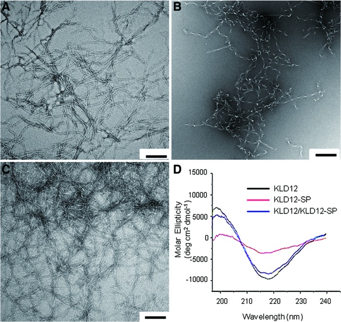
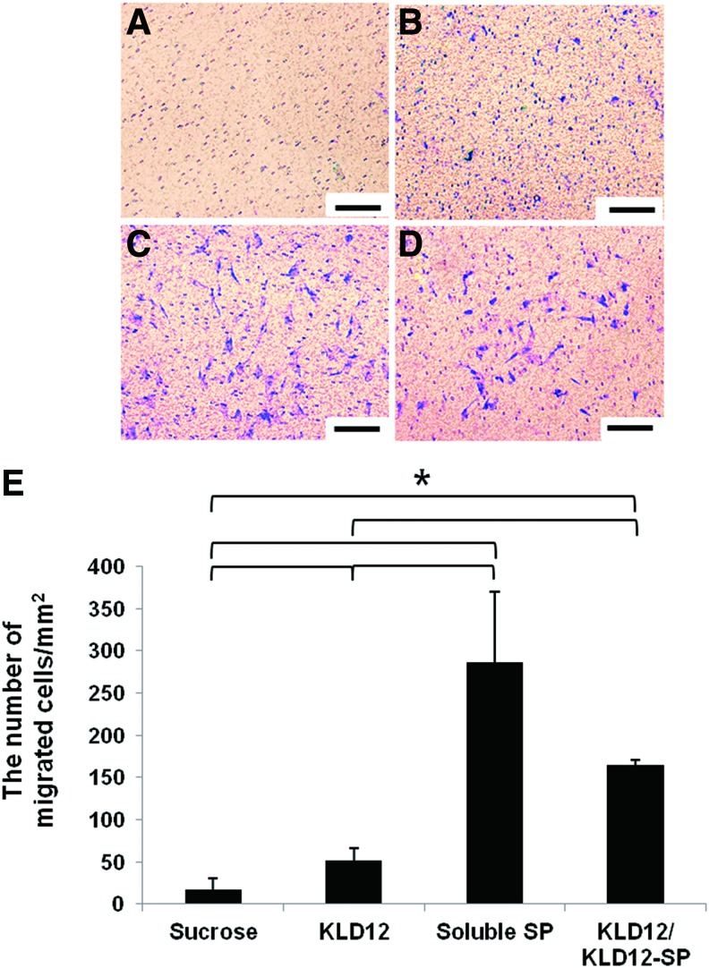
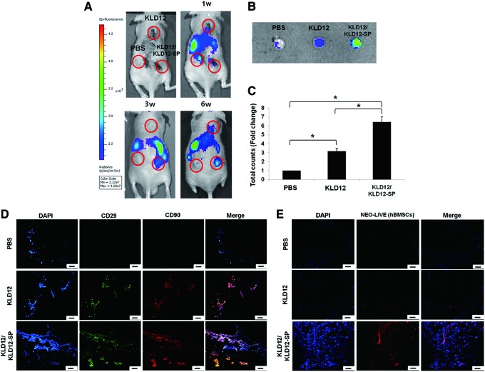
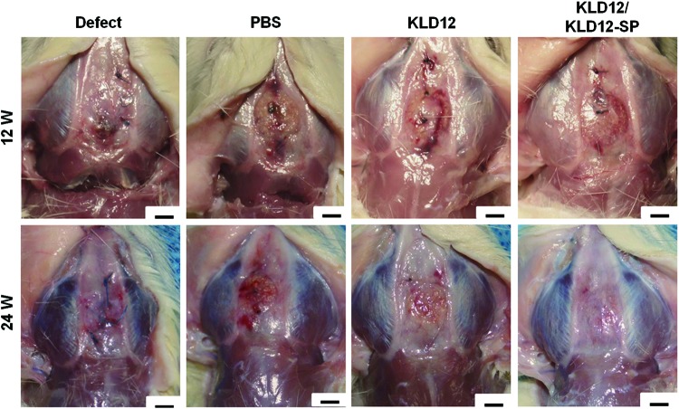
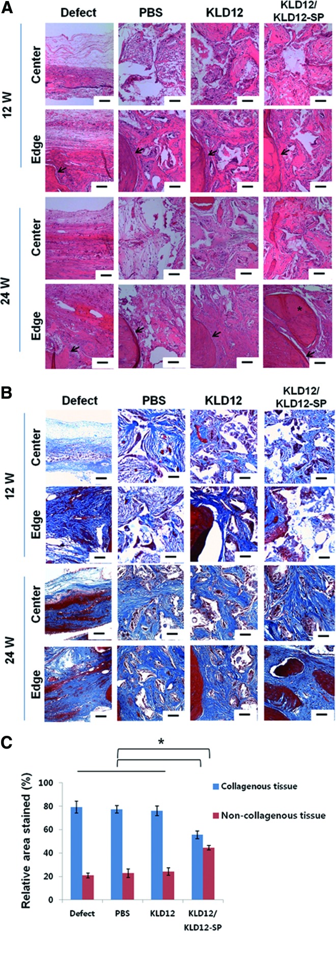
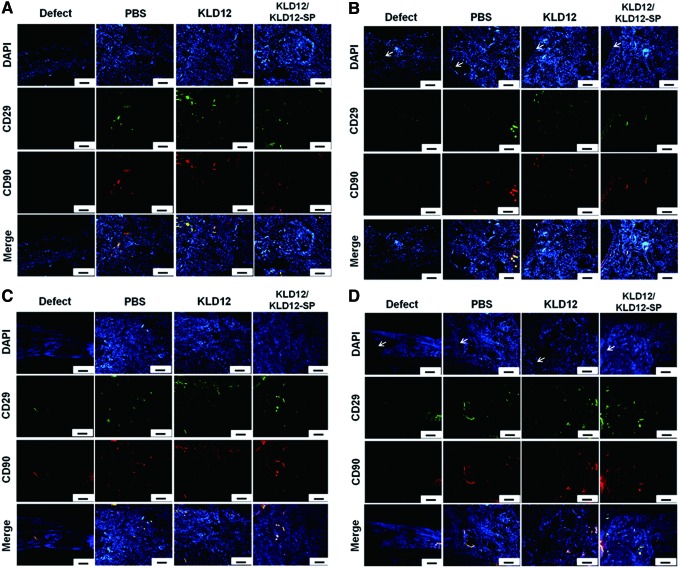
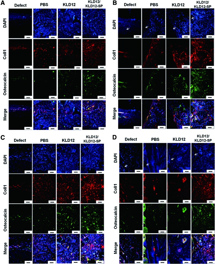
The number of patients requiring flat bone transplantation continues to increase worldwide. Cell transplantation has been successfully applied clinically; however, it causes another defect site and the time requirements to harvest cells and expand them are considerable. In this study, KLD12/KLD12-SP (KLD12+KLD12-substance P [SP]) was designed to mimic endogenous tissue-healing processes. The structures of KLD12, KLD12-SP, and KLD12/KLD12-SP were observed by transmission electron microscopy and circular dichroism spectra. KLD12/KLD12-SP nanofibers (5-10 nm) were created under physiological conditions by formation of a β-sheet structure. The ability of mesenchymal stem cells (MSCs) to recruit KLD12/KLD12-SP was observed by using an in vivo fluorescence imaging system. Labeled human bone marrow stromal cells supplied via an intravenous injection were recruited to the scaffold containing KLD12/KLD12-SP. Polylactic acid/beta-tricalcium phosphate (PLA/β-TCP) scaffolds filled with KLD12/KLD12-SP were applied to repair calvarial defects. The composite constructs (groups: defect, PLA/β-TCP, PLA/β-TCP/KLD12, and PLA/β-TCP/KLD12/KLD12-SP) were implanted into rat defect sites. Bone tissue regeneration was evaluated by observing gross morphology by hematoxylin and eosin and Masson's trichrome staining at 12 and 24 weeks after surgery. Gross morphology showed that the defect site was filled with new tissue that was integrated with host tissue in the KLD12/KLD12-SP group. In addition, from the staining data, cells were recruited to the defect site and lacunae structures formed in the KLD12/KLD12-SP group. From these results, the PLA/β-TCP+KLD12/KLD12-SP composite construct was considered for enhancement of bone tissue regeneration without cell transplantation.
Figures
Similar articles
-
Rat bone marrow stromal cells-seeded porous gelatin/tricalcium phosphate/oligomeric proanthocyanidins composite scaffold for bone repair.J Tissue Eng Regen Med. 2013 Sep;7(9):708-19. doi: 10.1002/term.1461. Epub 2012 Mar 6. J Tissue Eng Regen Med. 2013. PMID: 22392838
-
[An experimental study on repairing bone defect with composite of beta-tricalcium phosphate-hyaluronic acid-type I collagen-marrow stromal cells].Zhongguo Xiu Fu Chong Jian Wai Ke Za Zhi. 2005 Jun;19(6):468-72. Zhongguo Xiu Fu Chong Jian Wai Ke Za Zhi. 2005. PMID: 16038466 Chinese.
-
Tissue-engineered bone formation in vivo for artificial laminae of the vertebral arch using β-tricalcium phosphate bioceramics seeded with mesenchymal stem cells.Spine (Phila Pa 1976). 2013 Oct 1;38(21):E1300-6. doi: 10.1097/BRS.0b013e3182a3cbb3. Spine (Phila Pa 1976). 2013. PMID: 23873227
-
[Research progress of self-assembling peptide nanofiber scaffold for bone repair].Zhongguo Xiu Fu Chong Jian Wai Ke Za Zhi. 2014 Oct;28(10):1303-6. Zhongguo Xiu Fu Chong Jian Wai Ke Za Zhi. 2014. PMID: 25591312 Review. Chinese.
-
Intraoperative Construct Preparation: A Practical Route for Cell-Based Bone Regeneration.Tissue Eng Part B Rev. 2018 Oct;24(5):403-417. doi: 10.1089/ten.TEB.2018.0010. Epub 2018 May 9. Tissue Eng Part B Rev. 2018. PMID: 29631489 Review.
Cited by
-
No pain, no gain? The effects of pain-promoting neuropeptides and neurotrophins on fracture healing.Bone. 2020 Feb;131:115109. doi: 10.1016/j.bone.2019.115109. Epub 2019 Nov 9. Bone. 2020. PMID: 31715336 Free PMC article. Review.
-
Enzyme-responsive progelator cyclic peptides for minimally invasive delivery to the heart post-myocardial infarction.Nat Commun. 2019 Apr 15;10(1):1735. doi: 10.1038/s41467-019-09587-y. Nat Commun. 2019. PMID: 30988291 Free PMC article.
-
Self-assemble peptide biomaterials and their biomedical applications.Bioact Mater. 2019 Feb 13;4:120-131. doi: 10.1016/j.bioactmat.2019.01.002. eCollection 2019 Dec. Bioact Mater. 2019. PMID: 31667440 Free PMC article. Review.
-
A Procedural Overview of the Involvement of Small Molecules in the Nervous System in the Regulation of Bone Healing.Int J Nanomedicine. 2025 Jan 31;20:1263-1284. doi: 10.2147/IJN.S505677. eCollection 2025. Int J Nanomedicine. 2025. PMID: 39906525 Free PMC article. Review.
-
Neuro-bone tissue engineering: emerging mechanisms, potential strategies, and current challenges.Bone Res. 2023 Dec 20;11(1):65. doi: 10.1038/s41413-023-00302-8. Bone Res. 2023. PMID: 38123549 Free PMC article. Review.
References
-
- Szpalski C., Barr J., Wetterau M., Saadeh P.B., and Warren S.M.Cranial bone defects: current and future strategies. Neurosurg Focus 29,E8, 2010 - PubMed
-
- Elsalanty M.E., Por Y.C., Genecov D.G., Salyer K.E., Wang Q., Barcelo C.R., Troxler K., Gendler E., and Opperman L.A.Recombinant human BMP-2 enhances the effects of materials used for reconstruction of large cranial defects. J Oral Maxillofac Surg 66,277, 2008 - PubMed
-
- Jung Y., Kim S.S., Kim Y.H., Kim S.H., Kim B.S., Kim S., and Choi C.Y.A poly(lactic acid)/calcium metaphosphate composite for bone tissue engineering. Biomaterials 26,6314, 2005 - PubMed
-
- Cowan C.M., Shi Y.Y., Aalami O.O., Chou Y.F., Mari C., Thomas R., Quarto N., Contag C.H., Wu B., and Longaker M.T.Adipose-derived adult stromal cells heal critical-size mouse calvarial defects. Nat Biotechnol 22,560, 2004 - PubMed
-
- Zou D., Zhang Z., Ye D., Tang A., Deng L., Han W., Zhao J., Wang S., Zhang W., Zhu C., Zhou J., He J., Wang Y., Xu F., Huang Y., and Jiang X.Repair of critical-sized rat calvarial defects using genetically engineered bone marrow-derived mesenchymal stem cells overexpressing hypoxia-inducible factor-1alpha. Stem Cells 29,1380, 2011 - PubMed
Publication types
MeSH terms
Substances
LinkOut - more resources
Full Text Sources
Other Literature Sources
