. 2014 Nov 20;8(11):e3295.
doi: 10.1371/journal.pntd.0003295. eCollection 2014.

Pathophysiologic and transcriptomic analyses of viscerotropic yellow fever in a rhesus macaque model

Affiliations

Pathophysiologic and transcriptomic analyses of viscerotropic yellow fever in a rhesus macaque model

Flora Engelmann et al. PLoS Negl Trop Dis. .

Abstract

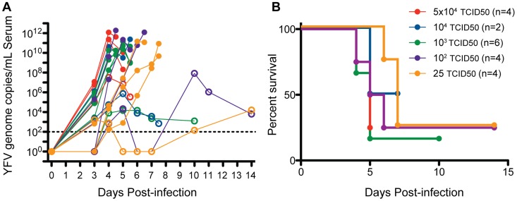
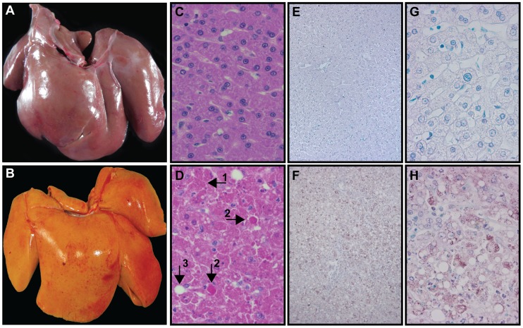
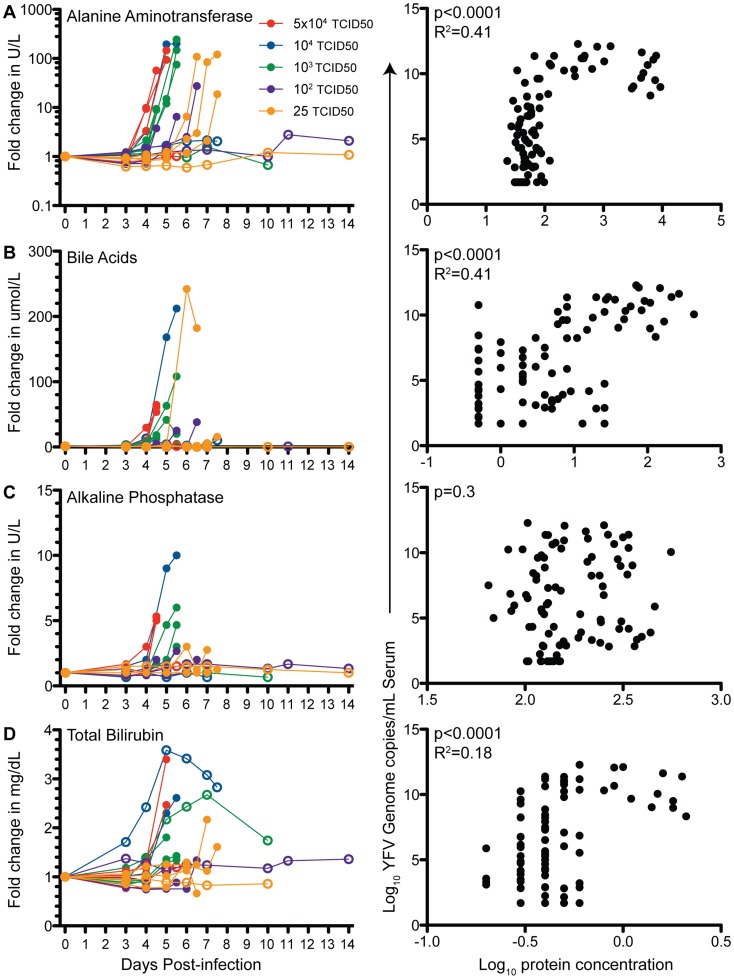
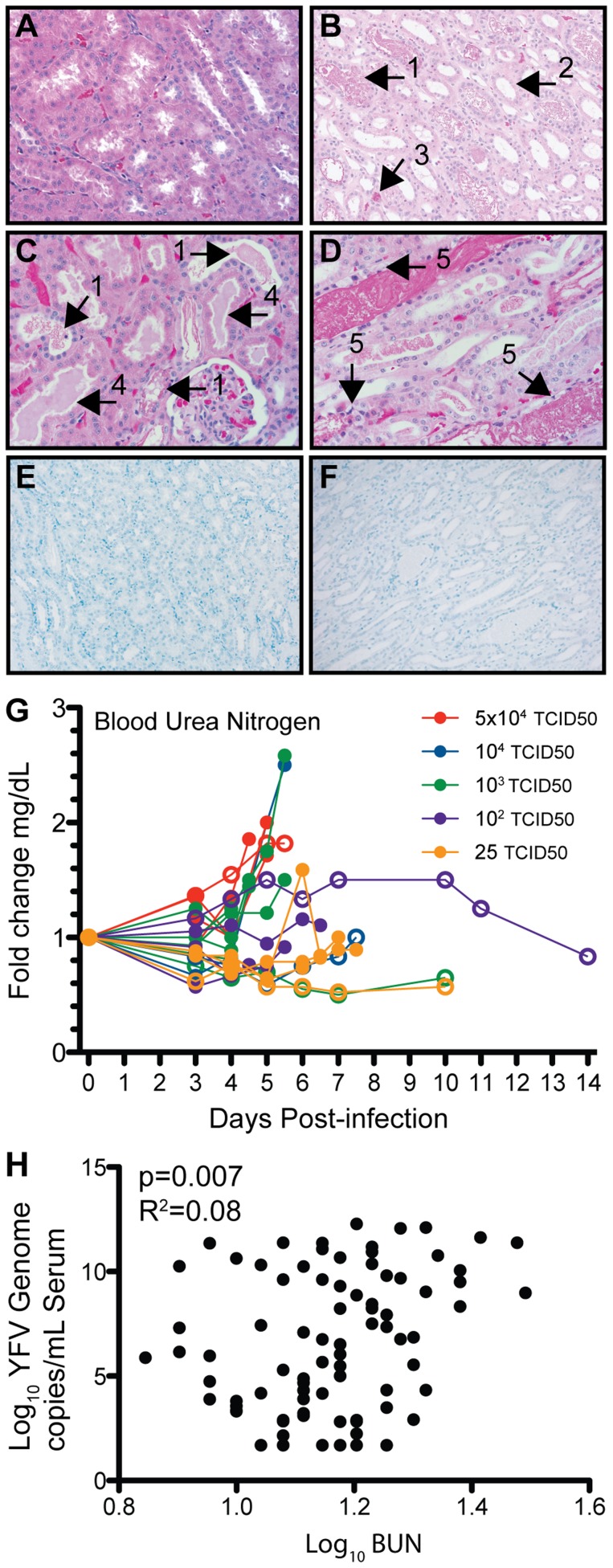
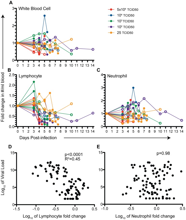
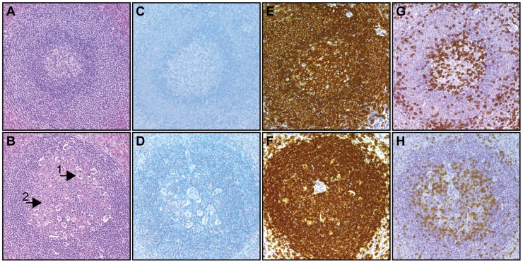
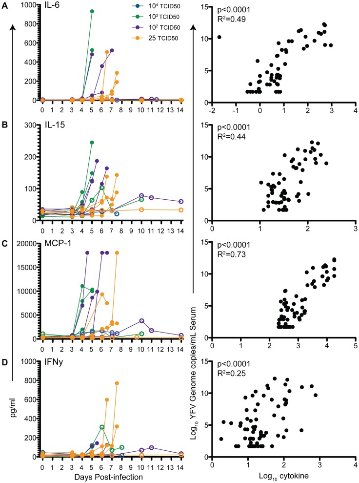
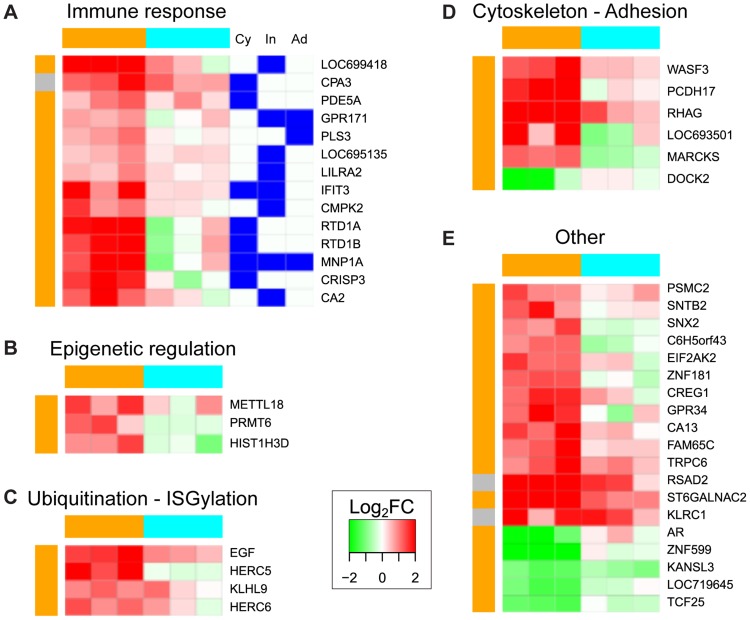
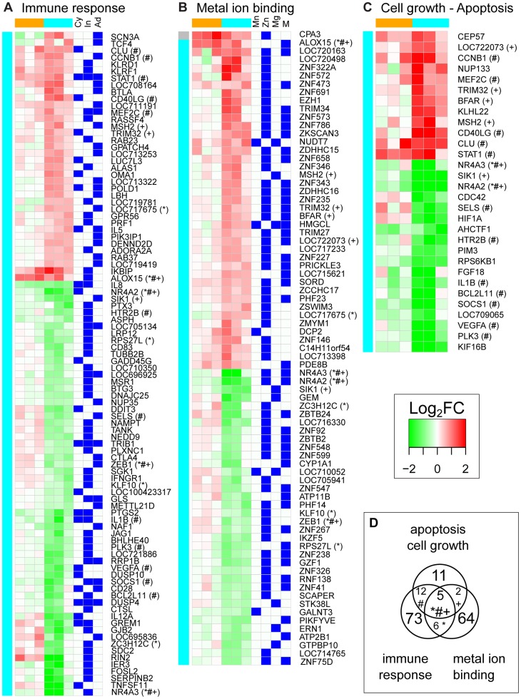
Infection with yellow fever virus (YFV), an explosively replicating flavivirus, results in viral hemorrhagic disease characterized by cardiovascular shock and multi-organ failure. Unvaccinated populations experience 20 to 50% fatality. Few studies have examined the pathophysiological changes that occur in humans during YFV infection due to the sporadic nature and remote locations of outbreaks. Rhesus macaques are highly susceptible to YFV infection, providing a robust animal model to investigate host-pathogen interactions. In this study, we characterized disease progression as well as alterations in immune system homeostasis, cytokine production and gene expression in rhesus macaques infected with the virulent YFV strain DakH1279 (YFV-DakH1279). Following infection, YFV-DakH1279 replicated to high titers resulting in viscerotropic disease with ∼72% mortality. Data presented in this manuscript demonstrate for the first time that lethal YFV infection results in profound lymphopenia that precedes the hallmark changes in liver enzymes and that although tissue damage was noted in liver, kidneys, and lymphoid tissues, viral antigen was only detected in the liver. These observations suggest that additional tissue damage could be due to indirect effects of viral replication. Indeed, circulating levels of several cytokines peaked shortly before euthanasia. Our study also includes the first description of YFV-DakH1279-induced changes in gene expression within peripheral blood mononuclear cells 3 days post-infection prior to any clinical signs. These data show that infection with wild type YFV-DakH1279 or live-attenuated vaccine strain YFV-17D, resulted in 765 and 46 differentially expressed genes (DEGs), respectively. DEGs detected after YFV-17D infection were mostly associated with innate immunity, whereas YFV-DakH1279 infection resulted in dysregulation of genes associated with the development of immune response, ion metabolism, and apoptosis. Therefore, WT-YFV infection is associated with significant changes in gene expression that are detectable before the onset of clinical symptoms and may influence disease progression and outcome of infection.


Conflict of interest statement

Oregon Health and Science University, MKS, and EH declare a financial interest based on shares in Najít Technologies, Inc., a company that may have a commercial interest in the results of this research and technology. This potential individual and institutional conflict of interest has been reviewed and managed by Oregon Health and Science University. This does not alter our adherence to all PLOS policies on sharing data and materials.

Figures

Figure 1
Figure 1. Wild type yellow fever virus (YFV-DakH1279) is highly virulent in rhesus macaques.
Twenty adult female rhesus macaque (8–16 years) were infected subcutaneously with YFV-DakH1279 strain with doses ranging from 25 to 5×10⁴ infectious units. (A) Viral loads were determined using quantitative RT-PCR and are expressed as genome copy number/ml serum. Filled circles denote animals that required humane euthanasia and open circles denote animals that survived YFV infection. (B) Kaplan-Meier survival curves following YFV-DakH1279 infection.
Figure 2
Figure 2. YFV-DakH1279 infection results in severe liver damage in rhesus macaques.
(A–B) Images of liver in a representative uninfected (A) and YFV-DakH1279 infected animal that required humane euthanasia (B). The infected liver is discolored with signs of hemorrhagic foci. (C–D, 400×) H&E staining of liver sections from an uninfected (C) and YFV-DakH1279-infected (D) animal. Extensive hepatocytes necrosis (1) along with eosinophilic degeneration of liver cells (Councilman bodies, 2), and fatty changes (3) are noted by the black arrows in panel D. (E–H) Histological analysis of YFV antigen in an animal that survived (E 100× & G 400×) or required humane euthanasia following YFV-DakH1279 infection (F 100× &H 400×).
Figure 3
Figure 3. Indices of liver injury in YFV-DakH1279-infected rhesus macaques.
Fold change in serum levels and correlation with viral genome copy number of alanine aminotransferase (A), bile acids (B), alkaline phosphatase (C) and total bilirubin (D) were determined at the indicated time points after infection. Filled circles denote animals that required humane euthanasia and open circles denote animals that survived YFV-DakH1279 infection.
Figure 4
Figure 4. Kidney injury in YFV-DakH1279-infected rhesus macaques.
(A–D) H&E staining of kidney sections from a representative uninfected (A) and YFV-DakH1279 infected animal (B–D). The black arrows note granular bilirubin casts in dilated distal convoluted tubules (1), necrotic tubular epithelial cells (2) and red blood cells (3), proteinaceous casts in dilated proximal convoluted tubules (4), and necrotic tubular epithelial cells in distal tubular segments containing large granular bilirubin casts (5). Histological staining of YFV antigen in kidney section from a surviving animal (E) and one that required euthanasia following YFV-DakH1279 infection (F) shows no detectable viral antigen. Slides A, C, and D acquired at 400× and slides B, E, and F acquired at 200× magnification. (G) Increased BUN levels were observed in some animals shortly before requiring euthanasia and (H) correlate with viral genome copy numbers/mL serum. Filled circles denote animals that required euthanasia and open circles denote animals that survived YFV infection.
Figure 5
Figure 5. YFV-DakH1279 infection results in severe lymphopenia.
(A) Fold change in white blood cells, (B) lymphocytes and (C) neutrophils were measured at the indicated time points and calculated as the ratio of cell counts/mL for each day relative to the 0 dpi count. (D) A negative correlation between viral load and lymphocyte count (p<0.0001, R2 = 0.46) (D), but no correlation was noted between numbers of circulating neutrophils and viral loads (E). Filled circles denote animals that required euthanasia and open circles denote animals that survived YFV infection.
Figure 6
Figure 6. Germinal center necrosis following infection with YFV-DakH1279.
(A, B) H&E staining showing healthy lymphocytic elements in the germinal centers of spleens from a surviving animal (A) and germinal necrosis in spleen of one that required euthanasia (B) Tangible body macrophage (1) and apoptosis (2) are indicated in panel B. (C, D) Histological examination of YFV antigen in spleen of a surviving animal (C) and one requiring euthanasia (D) show no viral antigen. (E–H) Histological staining for CD20 (E-survivor, F-euthanized) and CD3 antigens (G-survivor, H-euthanized). All slides were acquired at 200× magnification.
Figure 7
Figure 7. Circulating levels of inflammatory cytokines in YFV-DakH1279-infected rhesus macaques.
Serum levels of IL-6 (A), IL-15 (B), MCP-1 (C), and IFNγ (D) increase sharply in animals requiring euthanasia following infection with YFV and significantly correlate with viral loads. Filled circles denote animals that required euthanasia and open circles denote animals that survived YFV infection.
Figure 8
Figure 8. Functional characterization of PBMCs transcriptomic response to YFV-17D infection at 3 days post-infection.
The 46 DEGs after YFV-17D infection were grouped into functional categories based on enriched gene ontology (GO) terms (Table S1) and their expression value in log2FC is depicted in a green to red gradient color scheme. Three animals were challenged with YFV-17D (orange), while another three animals were infected with YFV-DakH1279 (cyan). Color on the left of each heatmap indicates whether the gene was found differentially expressed after YFV-17D infection (orange), or both YFV-17D and YFV-DakH1279 (grey). Genes were functionally categorized into: (A) immune response category (these genes were either involved in cytokine signaling pathways (Cy), or specifically up-regulated in innate immune cells (In) or adaptive immune cells (Ad)); (B) epigenetics; 9C) Ubiquitination-ISGylation; (D) cytoskeleton-adhesion; or (E) did not map to a specific functional category.
Figure 9
Figure 9. Functional characterization of PBMCs transcriptomic response to YFV-DakH1279 infection at 3 days post-infection.
Expression values for DEGs after YFV-DakH1279 infection within the 3 main enriched functional categories (Table S1) are depicted in a green to red gradient color scheme. Three animals were challenged with YFV-17D (orange), while another three animals were infected with YFV-DakH1279 (cyan). Color on the left of each heatmap indicates whether the gene was found differentially expressed after YFV-DakH1279 infection (cyan), or both YFV-17D and YFV-DakH1279 (grey). Genes were functionally categorized into: (A) the immune response category (these genes were either involved in cytokine signaling pathways (Cy), or specifically up-regulated in innate immune cells (In) or adaptive immune cells (Ad)); (B) Metal ion binding (manganese ion binding (Mn), zinc ion binding (Zn) and other metal ion binding (M)); or (C) cell growth and apoptosis. (D) Extent of overlap between the three functional categories: # indicates genes common between cell growth/apoptosis and “immune response” pathways; + indicates genes common to “cell growth/apoptosis” and “metal ion binding” pathways; * indicates genes common to “metal ion binding” and “immune response” pathways.

References

    1. Russell MN, Cetron MS, Eidex RB (2006) The US-Certified Yellow Fever Vaccination Center Registry: a tool for travelers, state health departments, and vaccine providers. J Travel Med 13: 48–49. - PubMed
    1. Bryant JE, Holmes EC, Barrett AD (2007) Out of Africa: a molecular perspective on the introduction of yellow fever virus into the Americas. PLoS Pathog 3: e75. - PMC - PubMed
    1. Tomori O (2004) Yellow fever: the recurring plague. Crit Rev Clin Lab Sci 41: 391–427. - PubMed
    1. Barrett AD, Higgs S (2007) Yellow fever: a disease that has yet to be conquered. Annu Rev Entomol 52: 209–229. - PubMed
    1. Paessler S, Walker DH (2013) Pathogenesis of the viral hemorrhagic fevers. Annu Rev Pathol 8: 411–440. - PubMed

Publication types

MeSH terms