. 2014 Nov 20;5(11):e1528.
doi: 10.1038/cddis.2014.489.

Characterization of novel markers of senescence and their prognostic potential in cancer

Affiliations

Characterization of novel markers of senescence and their prognostic potential in cancer

M Althubiti et al. Cell Death Dis. .

Abstract

Cellular senescence is a terminal differentiation state that has been proposed to have a role in both tumour suppression and ageing. This view is supported by the fact that accumulation of senescent cells can be observed in response to oncogenic stress as well as a result of normal organismal ageing. Thus, identifying senescent cells in in vivo and in vitro has an important diagnostic and therapeutic potential. The molecular pathways involved in triggering and/or maintaining the senescent phenotype are not fully understood. As a consequence, the markers currently utilized to detect senescent cells are limited and lack specificity. In order to address this issue, we screened for plasma membrane-associated proteins that are preferentially expressed in senescent cells. We identified 107 proteins that could be potential markers of senescence and validated 10 of them (DEP1, NTAL, EBP50, STX4, VAMP3, ARMX3, B2MG, LANCL1, VPS26A and PLD3). We demonstrated that a combination of these proteins can be used to specifically recognize senescent cells in culture and in tissue samples and we developed a straightforward fluorescence-activated cell sorting-based detection approach using two of them (DEP1 and B2MG). Of note, we found that expression of several of these markers correlated with increased survival in different tumours, especially in breast cancer. Thus, our results could facilitate the study of senescence, define potential new effectors and modulators of this cellular mechanism and provide potential diagnostic and prognostic tools to be used clinically.


Figures

Figure 1
Figure 1
Analysis of the membrane faction of senescence EJp16 and EJp21. (a) Western blots of EJp16 and EJp21 without and with induced expression of exogenous p16 or p21, respectively, as determined by the presence of tet in the culture medium. (b) SA-β-Gal staining of EJp16 and EJp21 uninduced (Control) or 4 days after tet removal to induce the expression of exogenous p16 or p21 (Senescent). Blue staining and morphological changes are indicative of senescence. (c) Western blot analysis of lysates separated into cytosolic and membrane fractions of EJp21 and EJp16 uninduced (C) or 4 days after tet removal (S). Calnexin is used as a marker of membrane proteins and MAPK as a marker of the cytosolic fraction. (d) Number of membrane proteins differentially expressed in control and senescent EJp21 and EJp16, compared with those present in both conditions, together with a list of targets selected for validation, as determined by mass spectrometry
Figure 2
Figure 2
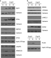
Western blot validation of senescent-specific targets in EJp16 and EJp21. (a and b) Protein expression of selected targets in the membrane fraction of lysates from EJp16 and EJp21 uninduced (C) or 4 days after tet removal (S). Calnexin and Na/K ATPase are used as membrane-specific loading controls
Figure 3
Figure 3
Expression of selected targets in membranes of senescent cells by cell fractionation. In all, 10–50% sucrose density gradient separation of lysates from EJp16, 4 days after tet removal. Calnexin and Na/K ATPase are used as markers of the cell membrane fractions. HDAC1 is used as marker of the nuclear fraction. MAPK is used as marker of the cytosolic fractions. SOD is used as marker of the mitochondrial fraction
Figure 4
Figure 4
Expression and localization of senescence markers. Immunofluorescent images of selected targets in EJp16 and EJp21 uninduced (Control) or 4 days after tet removal (Senescent), as well as early passage IMR90 human fibroblasts compared with those entering replicative senescence after serial passaging. Nucleus are stained with DAPI (blue)
Figure 5
Figure 5
Defining a new FACS-based protocol for the detection of senescent cells. (a) Representative plot analysis of fluorescence levels in control and senescent EJp16, HT1080p21-9 and human diploid fibroblasts (HDF) stained with fluorescently tagged antibodies against B2MG, DEP1 and NOTCH3, as measured by flow cytometry. Senescent cells were analysed after 5 days of p16 or p21 expression. Numbers indicate mean fluorescent intensity (MFI) values. (b) Average fold increases of MFI of the same cells when senescence is induced. Experiments were performed in triplicate. Error bars show S.D.
Figure 6
Figure 6
Expression of putative senescent markers in mouse and human tissues. Immunohistochemical staining of mouse (a) and human skin samples (b) with DEP1, NTAl, STX4 and B2MG antibodies. p16 is used as a known marker of senescence. Magnification: × 10 (mouse) and × 20 (human)
Figure 7
Figure 7
Correlation between senescent markers expression and survival in breast cancer. Kaplan–Meier survival curves of patients with breast cancer, segregated according to high (red) or low (green) expression of the genes from our panel of senescent markers, obtained from public databases through a bioinformatics analysis using PPISURV (www.bioprofiling.de). Each graph represents a different GEO data set

References

    1. Lowe SW, Cepero E, Evan G. Intrinsic tumour suppression. Nature. 2004;432:307–315. - PubMed
    1. Collado M, Serrano M. Senescence in tumours: evidence from mice and humans. Nat Rev Cancer. 2010;10:51–57. - PMC - PubMed
    1. Kuilman T, Michaloglou C, Mooi WJ, Peeper DS. The essence of senescence. Genes Dev. 2010;24:2463–2479. - PMC - PubMed
    1. Campisi J, d'Adda di Fagagna F. Cellular senescence: when bad things happen to good cells. Nat Rev Mol Cell Biol. 2007;8:729–740. - PubMed
    1. Dankort D, Filenova E, Collado M, Serrano M, Jones K, McMahon M. A new mouse model to explore the initiation, progression, and therapy of BRAFV600E-induced lung tumors. Genes Dev. 2007;21:379–384. - PMC - PubMed

Publication types

MeSH terms

Substances