. 2014 Nov 20;5(11):e1537.
doi: 10.1038/cddis.2014.501.

BIS targeting induces cellular senescence through the regulation of 14-3-3 zeta/STAT3/SKP2/p27 in glioblastoma cells

Affiliations

BIS targeting induces cellular senescence through the regulation of 14-3-3 zeta/STAT3/SKP2/p27 in glioblastoma cells

J-J Lee et al. Cell Death Dis. .

Abstract

Cellular senescence is an important mechanism for preventing tumor progression. The elevated expression of Bcl-2-interacting cell death suppressor (BIS), an anti-apoptotic and anti-stress protein, often correlates with poor prognosis in several cancers including glioblastoma; however, the role of BIS in the regulation of senescence has not been well defined. Here, we describe for the first time that the depletion of BIS induces G1 arrest and cellular senescence through the accumulation of p27 that is independent of p53, p21 or p16. The increase in p27 expression in BIS-depleted cells was attributable to an impairment of the ubiquitin-mediated degradation of p27, which was caused by a decrease in S-phase kinase-associated protein 2 (SKP2) at the transcriptional level. As an underlying molecular mechanism, we demonstrate that the loss of activity of signal transducer and activator of transcription 3 (STAT3) was specifically linked to the suppression of SKP2 expression. Despite a reduction in phospho-STAT3 levels, total STAT3 levels were unexpectedly increased by BIS depletion, specifically in the insoluble fraction. Our results show that 14-3-3ζ expression is decreased by BIS knockdown and that 14-3-3ζ depletion per se significantly induced senescence phenotypes. In addition, the ectopic expression of 14-3-3ζ blocked senescence caused by BIS depletion, which was paralleled with a decrease in insoluble STAT3 in A172 glioblastoma cells. These findings indicate that the impairment of the protein quality control conferred by BIS and/or 14-3-3ζ is critical for BIS depletion-induced senescence. Moreover, BIS knockdown also induced senescence along with an accumulation of total STAT3 and p27 in several different cell types as well as embryonic fibroblasts derived from Bis-knock out mice with/without variations in 14-3-3ζ levels. Therefore, our findings suggest that a downregulation of BIS expression could serve as a potential strategy for restricting tumor progression via an induction of senescence through the regulation of STAT3/SKP2/p27 pathway.


Figures

Figure 1
Figure 1
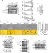
The depletion of BIS induces cell growth arrest and senescence in A172 glioblastoma cells. A172 cells were transfected with 100 nM of SiCON or SiBIS and incubated for the indicated times. d, day. (a) Western blot analysis for BIS expression. Actin levels are shown as a loading control. (b) Morphological changes and (c) SA-β-Gal staining and percentage (%) of SA-β-Gal-positive cells indicates that BIS depletion significantly induced senescence in A172 cells. ***P<0.001 versus day 0. Scale bars, 50 μm. (d) Cell viability was determined by Trypan blue staining. The cell number at day 0 was designated as 1.0. **P<0.01, ***P<0.001 versus control cells. (e) Colony-forming ability was compared between SiBIS- and SiCON-treated cells at 14 days following transfection. The relative efficiency was presented in right column. ***P<0.001 versus SiCON-treated cells. (f) Cell cycle distribution was measured by flow cytometry at 5 days following transfection. Values are mean±S.E. of triplicate experiments
Figure 2
Figure 2
p27 is essential for BIS knockdown-induced senescence independent of the p53 and p21 pathway. (a) Following SiBIS (100 nM) treatment for the indicated times, the expression levels of BIS, p53, p21, p27 and p-pRB were examined by western blotting. The expression levels of each protein from SiCON-treated cells were provided for comparison. (b) p27 protein expression accumulated with increasing doses of SiBIS, as determined at 5 days following transfection. The effect of BIS depletion on p27 expression and the induction of senescence was restored by the co-transfection of siRNA for p27 (Sip27, 100 nM) but not by siRNA for p53 (Sip53, 100 nM) as determined by western blotting (c), an observation for morphological changes (d) and SA-β-Gal staining and percentage (%) of SA-β-Gal-positive cells (e). Bars represent mean±S.E. of triplicate experiments. ***P<0.001. Scale bars, 50 μm
Figure 3
Figure 3
BIS modulates p27 levels via the regulation of 27 protein stability. (a) Relative mRNA levels for p27 were evaluated by qRT-PCR following SiCON or SiBIS (100 nM) treatment as described in the Materials and methods section. (b) p27 protein stability was examined by treatment of 40 μg/ml CHX for the indicated times at 24 h post-transfection of SiCON or SiBIS. p27 protein levels from each time point were normalized to β-actin protein levels with Image J software and are presented as the mean value±S.E. from three independent experiments (lower column). ***P<0.001 versus control cells. h, hour. (c) Treatment of MG132 (10 μM) for 1 h before CHX treatment for 2 h increased the stability of p27 in SiBIS-treated A172 cells. (d) The degradation of p27 caused by glucose (Glu)-free conditions was significantly prevented by BIS depletion. (e) The ubiquitination of p27 was decreased by BIS depletion. Two days following SiBIS transfection, cells were incubated in the presence or absence of glucose, and cell lysates were immunoprecipitated using p27 antibody or IgG, followed by immunoblotting with an ubiquitin (Ub) antibody. Total protein lysates were also subjected to western blot analyses with the indicated antibodies. (f) The interaction of endogenous BIS with p27 was investigated by immunoprecipitation of total cell lysates with BIS antibody and immunoblotted with p27 antibody
Figure 4
Figure 4
Inactivation of STAT3 is required for BIS knockdown-mediated senescence in A172 cells. (a) Immunoblotting for BIS, p27, SKP2, STAT3 and p-STAT3 (S727 and Y705) proteins in SiBIS (100 nM)-treated A172 cells. Densitometric analyses for each protein from three independent experiments were provided in lower column. ***P<0.001 versus the value from control cells. (b) Relative SKP2 mRNA levels were evaluated by qRT-PCR. Bars represent mean±S.E. from three independent experiments. ***P<0.001. (c)The effects of STAT3 phosphorylation status on BIS silencing-mediated morphological changes and SA-β-Gal activities were assessed by the transfection of 1 μg/ml of empty vector (HA), WT or STAT3 mutants (S727A and Y705F) concomitantly with SiCON or SiBIS (100 nM). Scale bars, 50 μm. (d) Reversal of SKP2 and p27 levels after WT-STAT3 was provided by western blot assay. Densitometric analysis for p27 from three independent experiments was shown in lower column. **P<0.01 and ***P<0.001 between indicated groups. (e) The effects of STAT3 depletion on STAT3, p-STAT3 (S727 and Y705), SKP2, p27, BIS and 14-3-3ζ status were examined by western blotting. (f) No significant decreases in p-JAK, p-mTOR or mTOR expression levels were observed following after SiBIS transfection
Figure 5
Figure 5
14-3-3ζ is involved in BIS depletion-induced senescence through the regulation of the STAT3 pathway. (a) Western blot assay for 14-3-3ζ and 14-3-3θ following SiBIS (100 nM) treatment. (b) The expression levels of 14-3-3ζ, p-STAT3 (S727 and Y705), STAT3, SKP2 and p27 were examined at the indicated times following 14-3-3ζ knockdown. (c) 14-3-3ζ depletion-induced senescence as determined by SA-β-Gal activities. ***P<0.001 versus control cells. (d) The overexpression of 14-3-3ζ prevented BIS depletion-induced senescence. Bars represent mean±S.E. from triplicate experiments.The percentage of SA-β-Gal-positive cells is shown in the right column. ***P<0.001 versus SiBIS-only treated cells. Scale bars, 50 μm. (e) Immunoprecipitation and immunoblotting was performed with the indicated antibodies following the transfection of STAT3, 14-3-3ζ or BIS-expressing plasmid for 2 days
Figure 6
Figure 6
BIS downregulation increases aggregated STAT3, which is restored by 14-3-3ζ. (a) STAT3 protein solubility is decreased by BIS knockdown (SiBIS, 100 nM). Total cell lysates were separated into soluble and insoluble fractions as described in Materials and methods section and subjected to a western blot assay for STAT3. (b) The effects of 14-3-3ζ on the solubility of STAT3 protein levels were examined by transfection with Myc-14-3-3ζ (1 μg/ml) with SiBIS and analyzed by western blot. The expression levels of BIS and 14-3-3ζ are verified in the right column. (c) Representative confocal immunofluorescence images analyzing endogenous STAT3 in A172 cells transfected with empty vector (Myc) or Myc-14-3-3ζ with SiCON or SiBIS. Endogenous STAT3 was detected using an anti-STAT3 antibody and FITC-conjugated anti-rabbit IgG. Lower panels represent higher magnifications of the selected area. Scale bars, 50 μm
Figure 7
Figure 7
Loss of BIS induces cellular senescence in various types of cells including Hep2, C6 and NMS. (a) Representative images of SA-β-Gal staining in each of the cell lines examined at 4 days following SiBIS transfection (100 nM). Scale bars, 50 μm. (b) Expression levels of BIS, 14-3-3ζ, p-STAT3 (S727 and Y705), STAT3, SKP2 and p27 were examined by immunoblotting from each of the cell lines
Figure 8
Figure 8
Serum limitation accelerates the induction of senescence in Bis-KO MEF cells. MEFs derived from Bis-WT or deficient mice were incubated with 10% FBS for 4 days. (a) Morphological changes and SA-β-Gal activities were evaluated. ***P<0.001. Scale bars, 50 μm. (b) Expression levels of p-STAT3, STAT3, p-pRB, p27 and 14-3-3ζ were determined by immunoblotting. (c) Skp2 transcript levels were determined by real-time PCR. ***P<0.001. (d) Proposed model summarizing how BIS depletion results in cellular senescence

References

    1. Schmitt CA. Senescence, apoptosis and therapy—cutting the lifelines of cancer. Nat Rev Cancer. 2003;3:286–295. - PubMed
    1. Nardella C, Clohessy JG, Alimonti A, Pandolfi PP. Pro-senescence therapy for cancer treatment. Nat Rev Cancer. 2011;11:503–511. - PubMed
    1. Kim WY, Sharpless NE. The regulation of INK4/ARF in cancer and aging. Cell. 2006;127:265–275. - PubMed
    1. Papazoglu C, Mills AA. p53: at the crossroad between cancer and ageing. J Pathol. 2007;211:124–133. - PubMed
    1. Lin HK, Chen Z, Wang G, Nardella C, Lee SW, Chan CH, et al. Skp2 targeting suppresses tumorigenesis by Arf-p53-independent cellular senescence. Nature. 2010;464:374–379. - PMC - PubMed

Publication types

MeSH terms