. 2014 Nov 20;10(11):e1004524.
doi: 10.1371/journal.ppat.1004524. eCollection 2014 Nov.

Human cytomegalovirus vaccine based on the envelope gH/gL pentamer complex

Affiliations

Human cytomegalovirus vaccine based on the envelope gH/gL pentamer complex

Felix Wussow et al. PLoS Pathog. .

Abstract

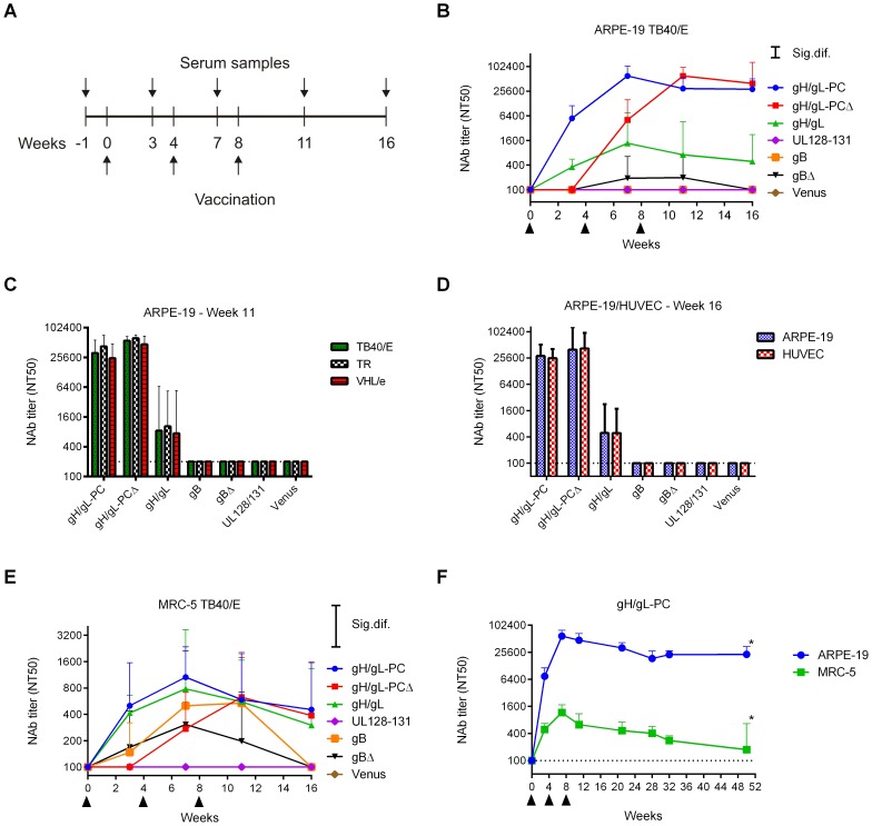
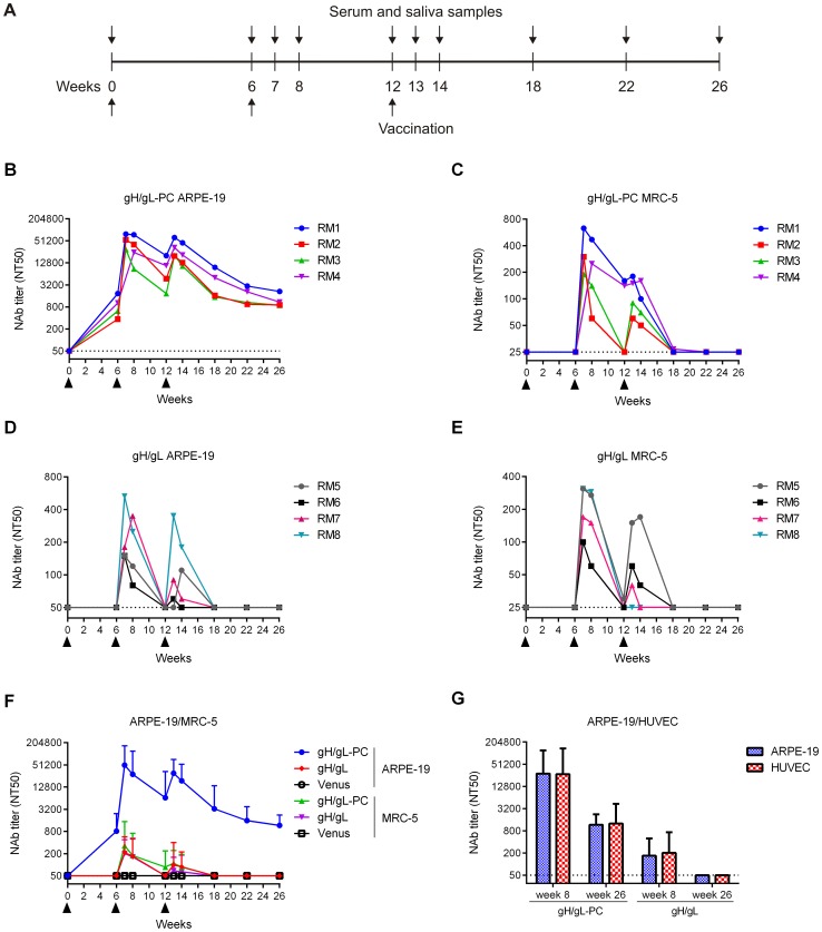
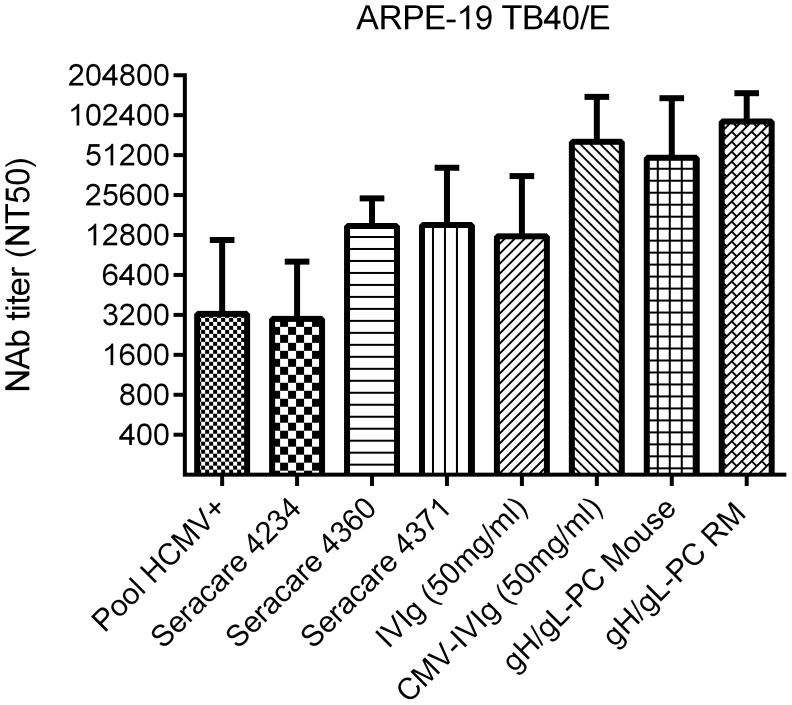
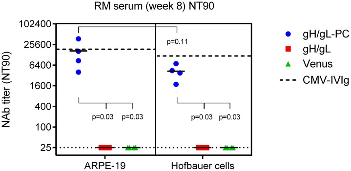
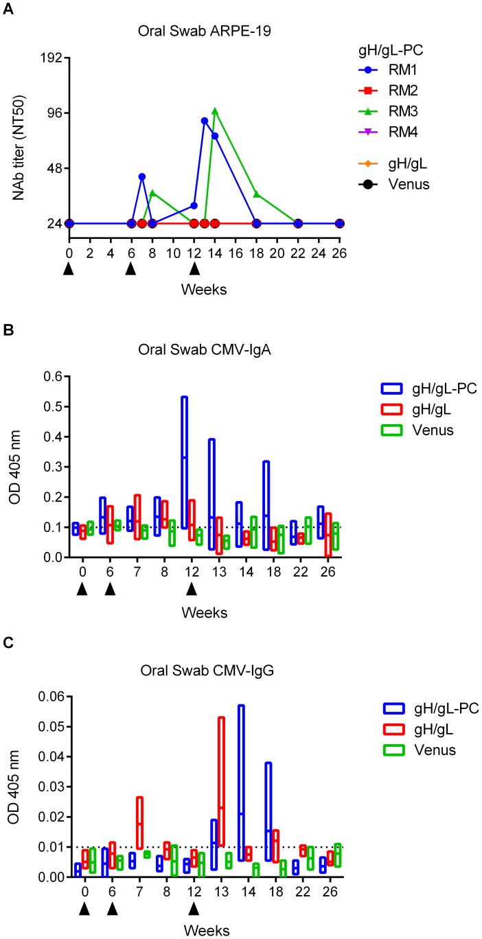
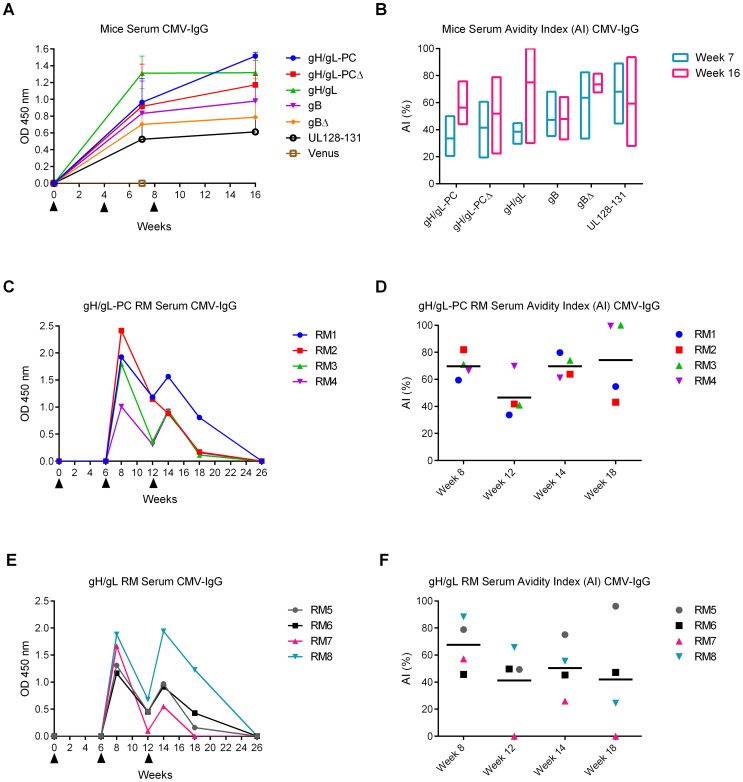
Human Cytomegalovirus (HCMV) utilizes two different pathways for host cell entry. HCMV entry into fibroblasts requires glycoproteins gB and gH/gL, whereas HCMV entry into epithelial and endothelial cells (EC) requires an additional complex composed of gH, gL, UL128, UL130, and UL131A, referred to as the gH/gL-pentamer complex (gH/gL-PC). While there are no established correlates of protection against HCMV, antibodies are thought to be important in controlling infection. Neutralizing antibodies (NAb) that prevent gH/gL-PC mediated entry into EC are candidates to be assessed for in vivo protective function. However, these potent NAb are predominantly directed against conformational epitopes derived from the assembled gH/gL-PC. To address these concerns, we constructed Modified Vaccinia Ankara (MVA) viruses co-expressing all five gH/gL-PC subunits (MVA-gH/gL-PC), subsets of gH/gL-PC subunits (gH/gL or UL128/UL130/UL131A), or the gB subunit from HCMV strain TB40/E. We provide evidence for cell surface expression and assembly of complexes expressing full-length gH or gB, or their secretion when the corresponding transmembrane domains are deleted. Mice or rhesus macaques (RM) were vaccinated three times with MVA recombinants and serum NAb titers that prevented 50% infection of human EC or fibroblasts by HCMV TB40/E were determined. NAb responses induced by MVA-gH/gL-PC blocked HCMV infection of EC with potencies that were two orders of magnitude greater than those induced by MVA expressing gH/gL, UL128-UL131A, or gB. In addition, MVA-gH/gL-PC induced NAb responses that were durable and efficacious to prevent HCMV infection of Hofbauer macrophages, a fetal-derived cell localized within the placenta. NAb were also detectable in saliva of vaccinated RM and reached serum peak levels comparable to NAb titers found in HCMV hyperimmune globulins. This vaccine based on a translational poxvirus platform co-delivers all five HCMV gH/gL-PC subunits to achieve robust humoral responses that neutralize HCMV infection of EC, placental macrophages and fibroblasts, properties of potential value in a prophylactic vaccine.


Conflict of interest statement

The authors have declared that no competing interests exist.

Figures

Figure 1
Figure 1. Insertion of gH/gL-PC subunits or gB into MVA-BAC.
A) Gene insertion using transfer vector. The transfer vector contains in the indicated order a mH5 promoter, a multiple cloning site (MCS), and a transcription termination signal (TS). It further comprises a I-SceI recognition site and a kanamycin resistance marker (KanR) both flanked by a 50 bp MVA sequence duplication (small black boxes). The entire construct is flanked by two ∼700 bp sequences of the MVA insertion site (large green and red bars). Following cloning, the gene is inserted into the MVA-BAC via a first Red recombination utilizing the large 700 bp sequence flanks. The selection marker is then removed after introduction of a double-strand break at the I-SceI site and a second Red recombination between the 50 bp sequence duplication. B) Gene insertion sites. HCMV genes were inserted with the indicated order (1-5) and 5′-3′ orientation into the MVA insertion sites Del2 (UL128), IGR3 (UL131), G1L/I8R (gH or gHΔTM, or at the ends of the BAC vector (B) within the Del3 insertion site (UL130 and gL). C) gB or gBΔTM were separately inserted into the G1L/I8R insertion site. Thin horizontal lines represent approximate lengths of either HCMV genes or portions of the MVA genome. Filled arrowheads in C indicate the cleavage site of the precursor gB protein encoded by the gB or gBΔTM gene cassette. AD1 designates antigenic domain 1, which is recognized by the anti-gB mAb 7-17.
Figure 2
Figure 2. Expression of gH/gL-PC subunits and gB from MVA.
Whole cell lysates of BHK cells or whole cell lysates and concentrated serum-free medium of CEF cells infected with the indicated MVA recombinants were analyzed by Immunoblot. gH/gL-PC subunits were detected with anti-HCMV gH mAb 11-1-1 and rabbit polyclonal antisera to UL128, UL130, UL131A, and gL. gB proteins were detected with anti-gB HCMV mAb 7-17, recognizing the C-terminal fragment of gB (Figure 1C). A and B) Immunoblot detection of gH/gL-PC subunits (A) or gB proteins (B) in MVA-infected BHK cells. C and D) Immunoblot detection of gH/gL-PC subunits (C) or gB proteins (D) in cell lysates (Cell) and concentrated medium (Med) of CEF cells infected with the MVA recombinants. MVA expressing the fluorescent marker Venus was a control (ctrl.) in A-D. Vaccinia virus BR5 was detected in the samples as loading control. PC  =  precursor protein; CT  =  C-terminal cleavage product of gB.
Figure 3
Figure 3. Complex formation of gH/gL-PC subunits co-expressed from MVA.
Cell lysates of BHK cells (prepared under non-denaturing conditions) or concentrated serum-free medium of CEF infected with MVA recombinants were subjected to co-IP with anti-HCMV gH mAb 11-1-1 or UL130 polyclonal antiserum. Immunoprecipitated gH/gL-PC subunits were detected with anti-gH mAb 11-1-1 and UL128, UL130, UL131A, and gL rabbit polyclonal antisera. A and B) Immunoblot detection of gH/gL-PC subunits after co-IP from BHK cells infected with MVA-gH/gL-PC, MVA-gH/gL, or MVA-UL128-131. C and D) Immunoblot detection of gH/gL-PC subunits following co-IP from concentrated medium of MVA-gH/gL-PCΔ-infected CEF cells. Irrelevant mouse IgG Ab and rabbit UL130 pre-immune serum were used for IP controls (ctrl.). IgGHC or IgGLC  =  Immunoglobulin G heavy or light chains. Input in A-D  =  detection of gH/gL-PC subunits in the samples subjected to co-IP.
Figure 4
Figure 4. Cell surface detection of gH and UL130 expressed from MVA recombinants.
BHK cells infected with MVA recombinants were analyzed by FC for cell surface staining of gH or UL130. Staining was performed with mouse anti-HCMV gH mAb 14-4b or UL130 rabbit antiserum as primary antibodies and fluorophore-coupled secondary Ab anti-mouse or anti-rabbit Alexa Fluor 647. A) FC analysis of cell surface staining of gH on BHK cells infected with the indicated gH/gL-PC-related vectors. B) FC analysis of cell surface staining of UL130 on BHK cells infected with the indicated gH/gL-PC-related vectors. Cells infected with MVA-Venus were analyzed as a control.
Figure 5
Figure 5. NAb induction by MVA recombinants in mice.
Balb/C mice were vaccinated three times by intraperitoneal (i.p.) route with MVA recombinants. HCMV-specific serum NAb titer (NT50) was determined at different time points post-vaccination. A) Schematic of time line for MVA vaccinations and serum sample collection. B-D) Shown are NT50 titers (geometric mean titer (GMT); n = 4) of vaccine groups measured at different time points on ARPE-19 cells against TB40/E (B), at week 11 on ARPE-19 cells against different HCMV strains (TB40/E, TR, VHL/e) (C), or at week 16 on ARPE-19 cells and HUVECs against TB40/E (D). E) NT50 titers measured on MRC-5 fibroblasts against TB40/E in the same samples as in B. F) Serum NT50 titers in MVA-gH/gL-PC-vaccinated mice (GMT; n = 8) measured on ARPE-19 and MRC-5 cells against TB40/E over one year (n = 4 at week 50 (asterisks)). Significance bars (Sig. dif.) in B and E indicate points of significance at p = 0.05. Dotted lines in all panels indicate the detection limit of the assay. Upper bars represent 95% confidence intervals.
Figure 6
Figure 6. HCMV-specific serum NAb titers in vaccinated RM.
Groups of four RM were vaccinated three times with MVA recombinants by intramuscular (i.m.) injection. HCMV-specific serum NT50 titers were determined at multiple time points on ARPE-19 cells, MRC-5 fibroblasts and HUVEC using HCMV TB40/E for infection. A) Schedule for vaccination and sample preparation. B-E) Serum NT50 titer of individual RM vaccinated with MVA-gH/gL-PC (B, C) or MVA-gH/gL (D, E) measured on ARPE-19 and MRC-5 cells. F) Comparison of serum NT50 titers determined in MVA-gH/gL-PC or MVA-gH/gL vaccine groups (GMT; n = 4) on ARPE-19 and MRC-5. Serum from RM vaccinated with MVA-Venus was analyzed as a control. G) Serum NT50 titer in vaccine groups (GMT, n = 4) measured on ARPE-19 cells and HUVECs at weeks 8 and 26 post-initial vaccination. Filled arrowheads indicate vaccinations. Dotted lines represent detection limits. Upper bars represent 95% confidence intervals.
Figure 7
Figure 7. NAb in vaccinated animals and HCMV-positive human sera.
NT50 titers were measured on ARPE-19 cells against TB40/E infection using commercially available HCMV IgG positive human sera (SeraCare Cat # 4234, 4360, 4371), HCMV pooled positive human sera (Pool HCMV+), IgG preparations (IVIg and CMV-IVIg), and in MVA-gH/gL-PC vaccinated animals after the first boost. Bars represent standard deviation of three independent experiments.
Figure 8
Figure 8. Neutralization of HCMV infection on placental macrophages (Hofbauer cells).
Serum samples of RM vaccinated with MVA-gH/gL-PC, MVA-gH/gL, or MVA-Venus from week 8 after initial vaccination (Figure 6A) were used to determine the titer that prevents 90% infection (NT90) of ARPE-19 cells and HC with HCMV strain TB40/E. Dashed lines indicate the NT90 values determined for HCMV-IVIg on ARPE-19 and Hofbauer cells. Dotted lines represent the detection limit. Statistical significance was evaluated using a two-sided Wilcoxon rank-sum test.
Figure 9
Figure 9. Ab response to HCMV in saliva of vaccinated RM.
Oral swab samples from RM vaccinated with MVA-gH/gL-PC, MVA-gH/gL, or MVA-Venus were analyzed for presence of NAb, HCMV mucosal IgA, and HCMV-IgG. A) NT50 titers in saliva of individual RM measured on ARPE-19 cells against TB40/E. B and C) HCMV IgG and IgA levels in saliva of vaccine groups (n = 4, analysis of single animals see Sup Figure 3). Floating bar charts represent time distribution of OD values obtained using HCMV-IgA or HCMV-IgG ELISA assay. Horizontal lines represent means; bars extend from minimum to the maximum values. Dotted lines represent 99% confidence interval upper limit for mean values obtained in sera from MVA-Venus RM. Filled triangles indicate time of MVA injections.
Figure 10
Figure 10. HCMV-specific Ab responses and avidity measurements in vaccinated animals.
HCMV IgG levels and avidity indices (AI) in serum samples of mice and RM were determined via ELISA using HCMV particles as antigen. A and B) HCMV IgG levels (GMT, n = 4) and AI measured in week 7 and 16 mouse serum samples. OD values at 450 nm were normalized to week zero OD. Floating bar chart in B represents the distribution of AI in different vaccine groups; bars extend from minimum to maximum values. C and E) HCMV IgG levels in individual RM vaccinated with MVA-gH/gL-PC (C) or MVA-gH/gL (E) using serum samples from different time points. D and F) HCMV-IgG AI values in individual RM vaccinated with MVA-gH/gL-PC or MVA-gH/gL determined at indicated time points. Horizontal bars represent group means. Filled triangles in A, C and E indicate MVA vaccinations.

References

    1. Plotkin SA (1999) Vaccination against cytomegalovirus, the changeling demon. Pediatr Infect Dis J 18: 313–325. - PubMed
    1. Weller TH (1971) The cytomegaloviruses: ubiquitous agents with protean clinical manifestations. II. N Engl J Med 285: 267–274. - PubMed
    1. Pereira L, Maidji E (2008) Cytomegalovirus infection in the human placenta: maternal immunity and developmentally regulated receptors on trophoblasts converge. Curr Top Microbiol Immunol 325: 383–395. - PubMed
    1. Griffiths P, Plotkin S, Mocarski E, Pass R, Schleiss M, Krause P, Bialek S (2013) Desirability and feasibility of a vaccine against cytomegalovirus. Vaccine 31 Suppl 2: B197–B203. - PMC - PubMed
    1. Schleiss MR (2013) Cytomegalovirus in the Neonate: Immune Correlates of Infection and Protection. Clin Dev Immunol 2013: 501801. - PMC - PubMed

Publication types

MeSH terms