Review
. 2015 May;28(5):612-30.
doi: 10.1038/modpathol.2014.158. Epub 2014 Nov 21.

Update for the practicing pathologist: The International Consultation On Urologic Disease-European association of urology consultation on bladder cancer

Affiliations
Review

Update for the practicing pathologist: The International Consultation On Urologic Disease-European association of urology consultation on bladder cancer

Mahul B Amin et al. Mod Pathol. 2015 May.

Abstract

The International Consultations on Urological Diseases are international consensus meetings, supported by the World Health Organization and the Union Internationale Contre le Cancer, which have occurred since 1981. Each consultation has the goal of convening experts to review data and provide evidence-based recommendations to improve practice. In 2012, the selected subject was bladder cancer, a disease which remains a major public health problem with little improvement in many years. The proceedings of the 2nd International Consultation on Bladder Cancer, which included a 'Pathology of Bladder Cancer Work Group,' have recently been published; herein, we provide a summary of developments and consensus relevant to the practicing pathologist. Although the published proceedings have tackled a comprehensive set of issues regarding the pathology of bladder cancer, this update summarizes the recommendations regarding selected issues for the practicing pathologist. These include guidelines for classification and grading of urothelial neoplasia, with particular emphasis on the approach to inverted lesions, the handling of incipient papillary lesions frequently seen during surveillance of bladder cancer patients, descriptions of newer variants, and terminology for urine cytology reporting.


Conflict of interest statement

Disclosure/conflict of interest The authors declare no conflict of interest.

Figures

Figure 1
Figure 1
Inverted papilloma and inverted papillary urothelial neoplasm of low malignant potential (PUNLMP). (a) An inverted urothelial papilloma shows endophytic growth of non-hyperplastic, non-atypical urothelium. (b) Often a suggestion of peripheral palisading is apparent, while the epithelium may frequently take on a bland, spindled appearance. (c) An inverted PUNLMP, similar to exophytic PUNLMP is composed of a hyperplastic (increased cells per unit area and/or increased thickness) urothelium growing in an endophytic pattern. (d) By definition, PUNLMP demonstrates no more than mild atypia and rare mitoses within a urothelium with preserved polarity.
Figure 2
Figure 2
Inverted papillary urothelial carcinoma, low-grade and high-grade. (a) Papillary urothelial carcinoma, low-grade, with predominant inverted growth shows a degree of cellularity and loss of polarity beyond that allowable in a PUNLMP. (b) Distinct, mild to moderate cytologic atypia is apparent. (c) Papillary urothelial carcinoma, high-grade, with predominant inverted growth shows even greater loss of order in the epithelium. This example showed foci of lamina propria invasion (asterisked), more extensive in adjacent fields, illustrated here to mainly contrast with the predominantly non-invasive component. (d) Loss of polarization with respect to the basement membrane is increased, while greater nuclear atypia is apparent; an atypical mitosis is identified.
Figure 3
Figure 3
Update on ‘formes frustes’ of papillary neoplasia. (a and b) Two examples of lesions that, if encountered in a biopsy of a patient under surveillance for papillary urothelial neoplasia, without a clinical impression of a papillary lesion may be termed ‘urothelial dysplasia with early papillary formations.’ Correlation with cystoscopic impression is key, as a lesion such as (b) may be diagnosed outright as papillary carcinoma, low-grade if clinically documented as a tumor. (c and d) Two examples of lesions that, if encountered in a similar scenario would be termed ‘urothelial carcinoma in situ with early papillary formations’; the lesion in (d) is better developed such that it may be considered sufficient to diagnose papillary urothelial carcinoma, high-grade, particularly if there is any endoscopic suspicion for a lesion. The ICUD notes that clinicopathologic correlation is essential to use these diagnostic terms.
Figure 4
Figure 4
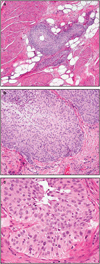
(a) The large nested variant of urothelial carcinoma may be high stage, as illustrated by deep muscularis propria invasion. (b) Despite the aggressive growth pattern, a well-polarized epithelium is preserved. (c) Atypia is less than expected for invasive carcinoma and raises consideration of low-grade inverted neoplasia.
Figure 5
Figure 5
Difficulties in diagnostic approach to large nested lesions. (a) This example of a muscularis propria invasive large nested urothelial carcinoma illustrates the diagnostic challenges, given cautery and crush artifacts and the low-grade appearance. If the lesions were not multifocally involving the muscularis propria, benign mimics such as urachal remnants or orifices of duplicated or tangentially sectioned ureters could be considered. (b) In another large nested case, the haphazard pattern of the nests is helpful in exclusion of benign anatomic or vestigial structures, as is their direct juxtaposition to large compact muscle bundles of muscularis propria (asterisked). Noninvasive inverted neoplasms should not generally extend into the muscularis propria. (c) Observation of a focus of conventional-type invasion (bracketed), consisting of irregularly sized and shaped invasive cell clusters, can be very helpful to exclude a non-invasive inverted neoplasm.
Figure 6
Figure 6
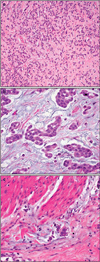
(a) Urothelial carcinoma with small tubules presents an infiltrative pattern of variably sized small tubules. (b) The epithelium lining the tubules is frequently attenuated, prompting consideration of nephrogenic adenoma or other processes. (c) These lesions may be deeply invasive of muscularis propria despite the low-grade appearance.
Figure 7
Figure 7
(a) Urothelial carcinoma with rhabdoid features is a pattern on the spectrum of poorly differentiated to undifferentiated urothelial carcinoma showing ‘rhabdoid’ morphology of discohe-sive cells with eccentric nuclei with prominent nucleoli and inclusion-like eosinophilic cytoplasmic inclusions. (b) Identification of a recognizable conventional urothelial carcinoma is helpful. (c) Expression of the urothelial carcinoma-associated marker, S100P, was diffuse, as were uroplakin II and GATA3 in this case.
Figure 8
Figure 8
(a) Urothelial carcinoma with chordoid features shows cords to reticular growth of epithelial cells in a myxoid stroma evocative of extraskeletal myxoid chondrosarcoma. (b) Another case shows clustered cells in abundant myxoid stroma. (c) These cases often present with high stage.
Figure 9
Figure 9
Update on features of micropapillary urothelial carcinoma. (a) Micropapillary urothelial carcinoma with invasion of the muscularis propria. (b) Prominent retraction artifact, apparent in this conventional urothelial carcinoma, may simulate micropapillary carcinoma. (c) Two specific features of micropapillary urothelial carcinoma illustrated here are ‘multiple nests in the same lacuna’ and ‘inverse polarization’ of the epithelium with peripherally oriented nuclei. (d) ‘Epithelial ring forms,’ asterisked at center, are another highly specific feature helpful in diagnosis.

References

    1. Kirkali Z, Chan T, Manoharan M, et al. Chapter 1. Bladder cancer: epidemiology, staging and grading, and diagnosis. In: Soloway M, Carmack A, Khoury S, editors. Bladder Tumors. 1st. Paris: Health Publications; 2005. pp. 13–64.
    1. Soloway M, Khoury S. Bladder Cancer. 2nd. Paris: 2012. EDITIONS 21.
    1. Amin MB, McKenney JK, Paner GP, et al. ICUD-EAU International Consultation on Bladder Cancer 2012: Pathology. Eur Urol. 2013;63:16–35. - PubMed
    1. Burger M, Oosterlinck W, Konety B, et al. ICUD-EAU International Consultation on Bladder Cancer 2012: Non-muscle-invasive urothelial carcinoma of the bladder. Eur Urol. 2013;63:36–44. - PubMed
    1. Palou J, Wood D, Bochner BH, et al. ICUD-EAU International Consultation on Bladder Cancer 2012: Urothelial carcinoma of the prostate. Eur Urol. 2013;63:81–87. - PubMed