

Next-generation sequencing of acute myeloid leukemia identifies the significance of TP53, U2AF1, ASXL1, and TET2 mutations
- PMID: 25412851
- PMCID: PMC5436901
- DOI: 10.1038/modpathol.2014.160
Next-generation sequencing of acute myeloid leukemia identifies the significance of TP53, U2AF1, ASXL1, and TET2 mutations
Abstract
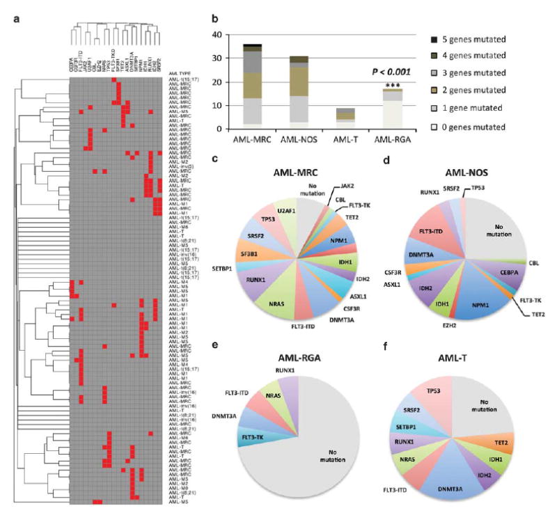
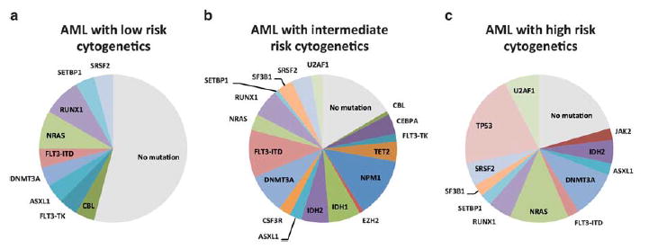
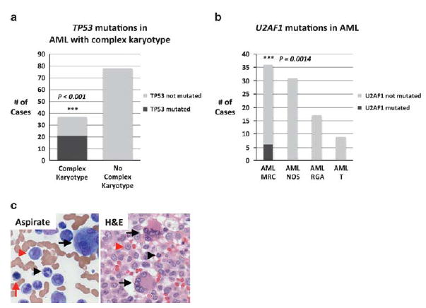
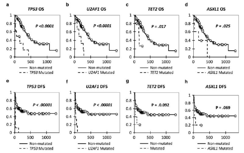
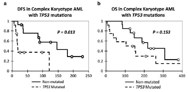
We assessed the frequency and clinicopathologic significance of 19 genes currently identified as significantly mutated in myeloid neoplasms, RUNX1, ASXL1, TET2, CEBPA, IDH1, IDH2, DNMT3A, FLT3, NPM1, TP53, NRAS, EZH2, CBL, U2AF1, SF3B1, SRSF2, JAK2, CSF3R, and SETBP1, across 93 cases of acute myeloid leukemia (AML) using capture target enrichment and next-generation sequencing. Of these cases, 79% showed at least one nonsynonymous mutation, and cases of AML with recurrent genetic abnormalities showed a lower frequency of mutations versus AML with myelodysplasia-related changes (P<0.001). Mutational analysis further demonstrated that TP53 mutations are associated with complex karyotype AML, whereas ASXL1 and U2AF1 mutations are associated with AML with myelodysplasia-related changes. Furthermore, U2AF1 mutations were specifically associated with trilineage morphologic dysplasia. Univariate analysis demonstrated that U2AF1 and TP53 mutations are associated with absence of clinical remission, poor overall survival (OS), and poor disease-free survival (DFS; P<0.0001), whereas TET2 and ASXL1 mutations are associated with poor OS (P<0.03). In multivariate analysis, U2AF1 and TP53 mutations retained independent prognostic significance in OS and DFS, respectively. Our results demonstrate unique relationships between mutations in AML, clinicopathologic prognosis, subtype categorization, and morphologic dysplasia.
Conflict of interest statement
Dr Robert Ohgami has been a consultant for Agilent Technologies. The other authors declare no conflict of interest.
Figures
References
-
- Vardiman JW, Thiele J, Arber DA, et al. The 2008 revision of the World Health Organization (WHO) classification of myeloid neoplasms and acute leukemia: rationale and important changes. Blood. 2009;114:937–951. - PubMed
-
- Visser O, Trama A, Maynadie M, et al. RARECARE Working Group. Incidence, survival and prevalence of myeloid malignancies in Europe. Eur J Cancer. 2012;48:3257–3266. - PubMed
-
- Sant M, Allemani C, Tereanu C, et al. HAEMACARE Working Group. Incidence of hematologic malignancies in Europe by morphologic subtype: results of the HAEMACARE project. Blood. 2010;116:3724–3734. - PubMed
-
- Deschler B, Lubbert M. Acute myeloid leukemia: epidemiology and etiology. Cancer. 2006;107:2099–2107. - PubMed
-
- Swerdlow SH, Campo E, Harris NL, et al. WHO Classification of Tumours of Haematopoietic and Lymphoid Tissues. 4. International Agency for Research on Cancer; Lyon, France: 2008. p. 439.
MeSH terms
Substances
Grants and funding
LinkOut - more resources
Full Text Sources
Other Literature Sources
Research Materials
Miscellaneous
