. 2014 Nov 21:5:5369.
doi: 10.1038/ncomms6369.

Divergent paths for the selection of immunodominant epitopes from distinct antigenic sources

Affiliations

Divergent paths for the selection of immunodominant epitopes from distinct antigenic sources

AeRyon Kim et al. Nat Commun. .

Abstract

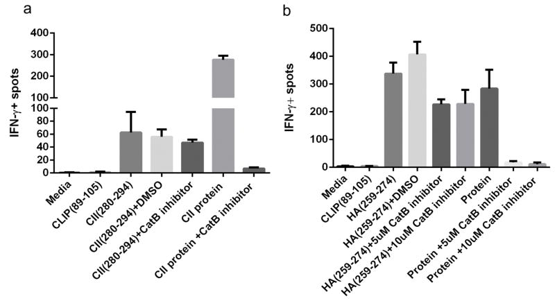
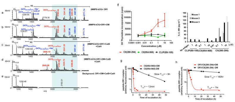
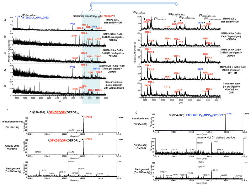
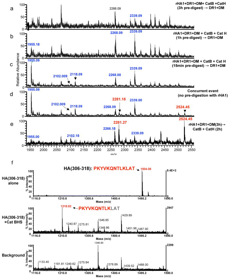
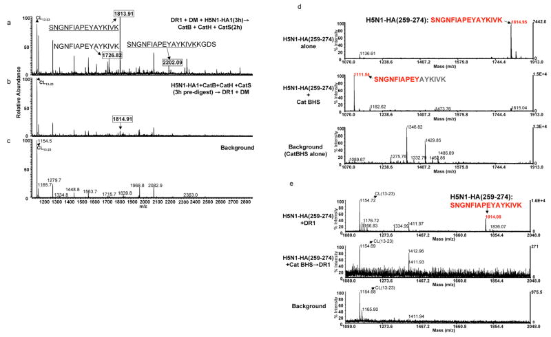
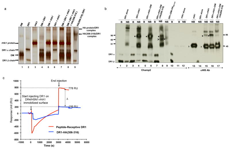
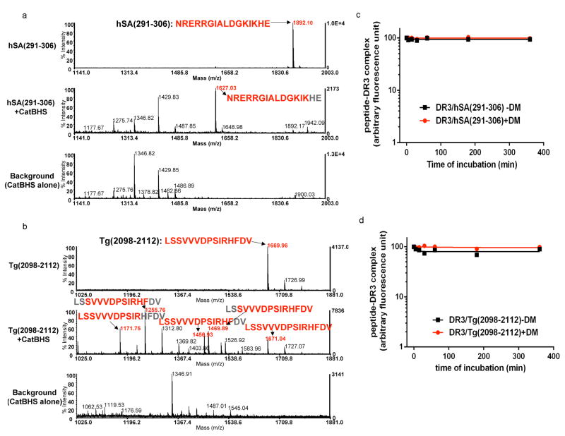
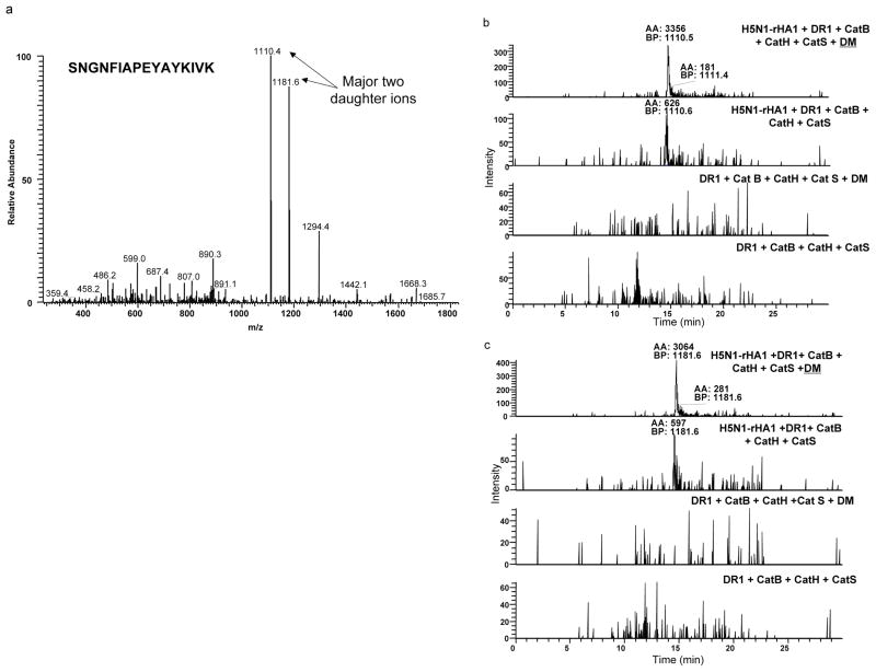
Immunodominant epitopes are few selected epitopes from complex antigens that initiate T-cell responses. Here to provide further insights into this process, we use a reductionist cell-free antigen-processing system composed of defined components. We use the system to characterize steps in antigen processing of pathogen-derived proteins or autoantigens and we find distinct paths for peptide processing and selection. Autoantigen-derived immunodominant epitopes are resistant to digestion by cathepsins, whereas pathogen-derived epitopes are sensitive. Sensitivity to cathepsins enforces capture of pathogen-derived epitopes by major histocompatibility complex class II (MHC class II) before processing, and resistance to HLA-DM-mediated-dissociation preserves the longevity of those epitopes. We show that immunodominance is established by higher relative abundance of the selected epitopes, which survive cathepsin digestion either by binding to MHC class II and resisting DM-mediated-dissociation, or being chemically resistant to cathepsins degradation. Non-dominant epitopes are sensitive to both DM and cathepsins and are destroyed.


Conflict of interest statement

Competing financial interest

The authors declare no competing financial interests.

Figures

Figure 1
Figure 1. Cathepsin B is a critical endosomal protease for generation of CII(280–294) and H5N1-HA(259–274) immunodominant epitope
ELISPOT assay measuring IFN-γ production of T cells isolated from DR1-transgenic mice immunized with CII(280–294) (a), or H5N1-HA(259–274) (b) in CFA. Cells were stimulated with peptides or proteins in vitro for 24h (a) or 48h (b) in the presence or absence of cell-permeable cathepsin B inhibitor, CA-074ME. Data shown here are representative of three independent experiments. Error bars are defined as SD.
Figure 2
Figure 2. Cathepsins and HLA-DM are necessary for the selection of the immunodominant epitope of type II collagen
(ad) Mass spectra of peptides eluted from DR1 containing MMP9-fragmented bCII. e shows the negative control reactions that do not contain the antigen. DR1 used in all experiments shown here (except for in sample d) was pre-incubated with HA(Y308A) to forms short-lived complexes with DR1(DR1/HA(Y308A) complex (T1/2 ~34 min) to induce a peptide-receptive conformation. The m/z 1258Da peak seen in e is background peptide peak an insect-derived protein that binds to a portion of purified DR1 and is present in most DR1 preparations. The peaks in the shaded area represent post-translationally modified variants of a dominant peptide composed of residues 273–305 of bCII (QTGEPGIAGFKGEQGPKGEPGPAGVQGAPGPAG). Mass species in red represent CII-derived peptides containing the immunodominant core CII(282–289). Non-dominant peptides are shown in blue. Background peptide species are labeled in black. These experiments were repeated more than three times and two cases all three cathepsins were included with similar results. (f) Proliferation of T cells isolated from DR1-transgenic mice immunized with bCII protein in CFA in response to stimulation with, CII(280–294), CII(954–968), or CLIP(89–105) in vitro. Cellular proliferation was measured by [3H] thymidine incorporation (Left panel). The data shown here is a representative result of one mouse out of four individual mice tested. Error bars are defined as SD. IL-2 ELISA was performed from supernatant collected from in vitro culture of another three individual mice immunized with CII protein/CFA. Cell culture supernatants were removed after 48h culture (Right panel). (g–h) Dissociation assay of fluorescently labeled non-dominant epitope, CII(954–968) and immunodominant epitope CII(280–294) from DR1. Fluorescently labeled CII(954–968)/DR1 complexes (g), or fluorescently labeled CII(280–294)/DR1 complexes (h) were dissociated in the presence of 100μM of unlabeled HA(306–318) at 37°C for the indicated times in the absence (square), or presence of DM (circle). The fluorescence of the labeled complex before dissociation is arbitrarily assigned a value of 100, and fluorescence after dissociation is expressed as a fraction of fluorescence before dissociation. Representative of three dissociation experiments.
Figure 3
Figure 3. Type II collagen-derived immunodominant peptides are resistant to cathepsin digestion
(a–e) Mass spectra of peptides eluted from DR1 are shown for m/z range of 1400–3600Da. (a) MMP9-bCII was incubated with DR1 and DM for 3h. (b) MMP9-bCII was first exposed to cathepsins B and H for 3h, (c) for 1h or (d) for 15min, and then incubated with DR1 and DM for additional 3h. (e) Mass spectrum of peptides eluted from DR1 when all components of the cell-free assay were mixed simultaneously and incubated together for 3h. As in Fig 1, the peaks in the shaded area between 3000–3550Da represent PTM variants of the dominant peptide bCII(273–305). Immunodominant peptides are labeled in red and non-dominant CII peptides are shown in blue. CII protein pre-digestion assays were repeated three times. (f) Mass spectra of synthetic CII(280–294) immunodominant peptide, and (g) Mass spectra of synthetic non-dominant CII(954–968) that were directly exposed to the cathepsins, B, H, and S for 1h at 37°C. Top spectra in both f and g show synthetic peptides without treatment. Middle spectra show samples incubated with the cathepsins. Bottom spectra correspond to the background samples containing only cathepsins. The peak at m/z 1338Da of the middle spectrum in (g) is not derived from the collagen CII(954–968) there is a background peptide from cathepsins. These experiments were repeated three times.
Figure 4
Figure 4. Degradation of HA(306–318) by the cathepsins outcompetes its capture by HLA-DR1
(a–e) The mass spectra of peptides eluted from DR1. Mass species in red represent rHA1 fragments containing the DR1 restricted immunodominant HA(306–318) epitope. Peptides highlighted in blue represent other rHA1-derived peptides eluted from DR1. Recombinant HA1 was first exposed to cathepsins B and H for (a) 3h, (b) 1h, or (c) 15 minutes, followed by incubation with DR1 and DM for 2 h at 37°C. (d) Mass spectrum of peptides eluted from DR1 when all components were mixed simultaneously and incubated together for 3h. (e) Mass spectra of peptides eluted from DR1 when rHA1 was incubated with DR1 and DM for 3h first, and then incubated with the cathepsins for additional 2h. (f) Sensitivity of synthetic HA(306–318) to the cathepsins tested by direct exposure to cathepsins B, H, and S. Top spectrum shows HA(306–318) peptide alone. Middle spectrum is the sample reaction containing HA(306–318) incubated with the cathepsins for 1h at 37°C, and the bottom spectrum shows the background sample containing the cathepsin mix without the synthetic peptide. Experiments were repeated three times.
Figure 5
Figure 5. Immunodominant epitope of H5N1-rHA1 is sensitive to the cathepsins
(a–c) The mass spectra of peptides eluted from DR1 when (a) denatured H5N1-rHA1 (A/Vietnam/1203/2004) was first incubated with DR1 and DM, and then exposed to cathepsins B, H, and S, or (b) when denatured H5N1-rHA1 was first exposed to the cathepsins for 3h and then incubated with DR1 and DM. (c) is the background spectrum. Boxed mass species represent H5N1-rHA1 fragments containing the DR1 restricted immunodominant HA(259–274) epitope. (d–e) mass spectra of the synthetic HA(259–274). (d) Similar to the procedure in Figure 3f, the top panel shows undigested HA(259–274) peptide. Digested peptide with the cathepsins is shown in the middle panel, and the bottom panel shows cathepsins alone as a background control. (e) Mass spectra of peptides eluted from DR1, after 3h incubation of DR1 with HA(259–274) peptide alone (top), HA(259–274) digested with the cathepsins (middle), or the cathepsins alone (bottom). Baculovirus-derived peptide CL(13–23) was detected in every spectra from samples containing DR1. Experiments were repeated three times.
Figure 6
Figure 6. Intact protein antigens form complexes with HLA-DR
(a–b) SDS-stable complex formation with intact proteins and DR1. Various combinations of DR1, DM, rHA1, and synthetic HA(306–318) peptide were incubated in citrate-phosphate buffer pH 5.0 for 2h at 37°C. Different combinations of proteins and peptides (shown in different lanes) were analyzed by “gentle” SDS-PAGE in which samples are not boiled prior to loading and (a) silver-stained, or (b) western blotted. Polyclonal anti-DR1 serum (CHAMP2) or anti-His antibody was used for detecting protein/DR complexes in (b). Recombinant HA1 protein/DR1 complexes (marked by *) migrated at approximately 96 kD molecular mass. Unbound rHA1 protein migrated at ~45kD (marked by 〉), HA(306–318)/DR1 migrated at around 60kD (marked by ➢), and DR1 is shown at about 50kD. B, boiled, NB, non-boiled.rHA1/DR1 complexes were estimated to migrate at 96 kD molecular mass in both a and b. (c) Simultaneous binding of two DR alleles to H5N1-rHA1 protein as detected by BIAcore. Biotinylated DR4/HA(Y308A) complex was incubated with denatured H5N1-rHA1 protein for 20min at 37°C in the presence of DM, during which HA(Y308A) would dissociate and be replaced by H5N1-HA1 protein. At the end of the incubation, DR4/H5N1-rHA1 protein complex was injected over the streptavidin coated sensor chip (SA) for 600 seconds and produced 3500 RU, shown as time zero. Then, receptive DR1 (pre-incubated with HA(Y308A) (red), or closed HA(306–318) complex (as control) (blue) were injected over H5N1-rHA1 bound DR4 immobilized surface in the presence of DM for 3000 seconds after which running buffer was injected over the chip to monitor the binding of proteins. Sensograms from both injections are superimposed in the figure for comparison. Negative sensograms observed during the injection are due to changes in buffer conditions between the running buffer and the sample buffer. Binding of the protein of interest to the immobilized protein are shown in the Response Unit (RU) indicating the RU difference between the injection start time versus injection end time.
Figure 7
Figure 7. HLA-DR3 binding core region of autoantigen derived immunodominant epitopes show resistance to the cathepsins and HLA-DM
(a–b) Mass spectra of retinal arrestin derived dominant epitope, hSA(291–306), and thyroglobulin-derived dominant epitope, Tg(2098–2112), after digestion with the cathepsins. Untreated synthetic peptides (a) hSA(291–306) or (b) Tg(2098–2112) are shown on the top spectra in a and b. Peptides were incubated with cathepsin B, H, and S for 1h at 37°C (shown in the middle spectra). Background controls that included cathepsins alone are shown in the bottom spectra. The experiments were repeated twice. (c–d) Dissociation kinetics of fluorescently labeled hSA(291–306) or Tg(2098–2112) from DR3. (c) FL-hSA(291–306)/DR3 or (d) FL-Tg(2098–2112)/DR3 complexes were formed over a three day incubation with peptides in 37°C and the unbound peptides were removed by spin column separation. Dissociation experiments were in the presence of 100μM unlabeled hSA(291–306) or 100μM unlabeled Tg(2098–2112) peptides at 37°C for the indicated time in the presence (circle) or absence (square) of DM. The fluorescence of the labeled complex before dissociation was arbitrarily assigned a value of 100.0, and fluorescence after dissociation is expressed as a fraction of fluorescence before dissociation. Dissociation experiments were repeated twice.
Figure 8
Figure 8. Repertoire of H5N1-rHA1 derived peptides appears the same with or without DM
Samples containing equal starting materials (same amount of DR1 and rHA1 protein) were prepared in the presence, or absence of DM and the eluted peptides were analyzed by MALDI. (a–d) Native form of H5N1-rHA1 protein, DR1, and the cathepsins B, and H, and S were incubated in the (a) presence or (b) absence of DM. (c) and (d) are background spectra for samples a and b, respectively, from reactions not containing H5N1-rHA1. Immunodominant epitopes, HA(259–274) (SNGNFIAPEYAYKIVK), and HA(259–278) (SNGNFIAPEYAYKIVKKGDS), are shown at m/z 1814.7Da and 2200.8Da. The experiments were repeated three times.
Figure 9
Figure 9. DM increases the abundance of dominant epitope
Quantitation ofHA(259–274) epitopes obtained in samples with or without DM by liquid chromatography (LC) combined with Linear Ion Trap Quadrouple tandem mass spectrometry (LC-LTQ MS/MS). Samples containing equal starting materials (same amount of DR1 and rHA1 protein) were prepared in the presence, or absence of DM and the eluted peptides were analyzed. For relative quantification of this peptide, the samples were rerun on LTQ mass spectrometry and HA(259–274) was detected as doubly charged ions at m/z 907 Da. (a) The MS/MS profile of the doubly charged ions produced two major daughter ions at m/z 1110 and 1181Da. (b) The base peak area values from extraction ion chromatograms of 1110 Da or (c) 1181 Da daughter ions in samples with or without DM are shown and were estimatedas 3356 and 3064 in the presence of DM, versus 626 and 597 without DM accordingly. Neither of those daughter ions were detected in the background samples. (BP -base peak; AA – automated area calculated using Genesis peak detection algorithm). The experiments were repeated twice and each experiment was quantified two times.

Similar articles

Cited by

References

    1. Vezys V, et al. Memory CD8 T-cell compartment grows in size with immunological experience. Nature. 2009;457:196–199. doi: 10.1038/nature07486. nature07486 [pii] - DOI - PubMed
    1. Sercarz EE, Maverakis E. Mhc-guided processing: binding of large antigen fragments. Nat Rev Immunol. 2003;3:621–629. - PubMed
    1. Dai G, Carmicle S, Steede NK, Landry SJ. Structural basis for helper T-cell and antibody epitope immunodominance in bacteriophage T4 Hsp10. Role of disordered loops. J Biol Chem. 2002;277:161–168. - PubMed
    1. Maric M, et al. Defective antigen processing in GILT-free mice. Science. 2001;294:1361–1365. - PubMed
    1. Delamarre L, Couture R, Mellman I, Trombetta ES. Enhancing immunogenicity by limiting susceptibility to lysosomal proteolysis. J Exp Med. 2006;203:2049–2055. - PMC - PubMed

Publication types

MeSH terms