

Human umbilical cord blood cells induce neuroprotective change in gene expression profile in neurons after ischemia through activation of Akt pathway
- PMID: 25413246
- PMCID: PMC4739737
- DOI: 10.3727/096368914X685311
Human umbilical cord blood cells induce neuroprotective change in gene expression profile in neurons after ischemia through activation of Akt pathway
Abstract
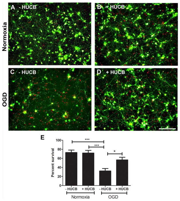
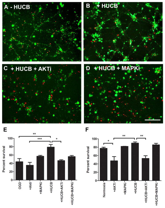
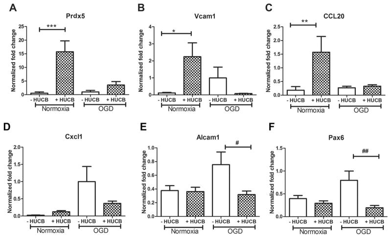
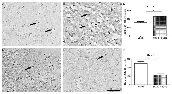
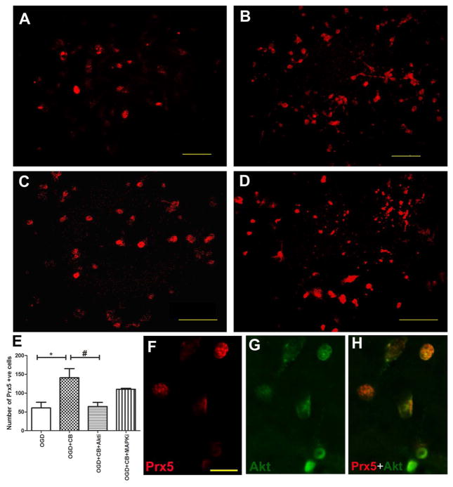
Human umbilical cord blood (HUCB) cell therapies have shown promising results in reducing brain infarct volume and most importantly in improving neurobehavioral function in rat permanent middle cerebral artery occlusion, a model of stroke. In this study, we examined the gene expression profile in neurons subjected to oxygen-glucose deprivation (OGD) with or without HUCB treatment and identified signaling pathways (Akt/MAPK) important in eliciting HUCB-mediated neuroprotective responses. Gene chip microarray analysis was performed using RNA samples extracted from the neuronal cell cultures from four experimental groups: normoxia, normoxia+HUCB, OGD, and OGD+HUCB. Both quantitative RT-PCR and immunohistochemistry were carried out to verify the microarray results. Using the Genomatix software program, promoter regions of selected genes were compared to reveal common transcription factor-binding sites and, subsequently, signal transduction pathways. Under OGD condition, HUCB cells significantly reduced neuronal loss from 68% to 44% [one-way ANOVA, F(3, 16)=11, p=0.0003]. Microarray analysis identified mRNA expression of Prdx5, Vcam1, CCL20, Alcam, and Pax6 as being significantly altered by HUCB cell treatment. Inhibition of the Akt pathway significantly abolished the neuroprotective effect of HUCB cells [one-way ANOVA, F(3, 11)=8.663, p=0.0031]. Our observations show that HUCB neuroprotection is dependent on the activation of the Akt signaling pathway that increases transcription of the Prdx5 gene. We concluded that HUCB cell therapy would be a promising treatment for stroke and other forms of brain injury by modifying acute gene expression to promote neural cell protection.
Figures
Similar articles
-
Neuroprotection by cord blood stem cells against glutamate-induced apoptosis is mediated by Akt pathway.Neurobiol Dis. 2008 Dec;32(3):486-98. doi: 10.1016/j.nbd.2008.09.005. Epub 2008 Sep 30. Neurobiol Dis. 2008. PMID: 18930139
-
Cord blood administration induces oligodendrocyte survival through alterations in gene expression.Brain Res. 2010 Dec 17;1366:172-88. doi: 10.1016/j.brainres.2010.09.078. Epub 2010 Sep 29. Brain Res. 2010. PMID: 20883670 Free PMC article.
-
Neuroprotective effects of pioglitazone in a rat model of permanent focal cerebral ischemia are associated with peroxisome proliferator-activated receptor gamma-mediated suppression of nuclear factor-κB signaling pathway.Neuroscience. 2011 Mar 10;176:381-95. doi: 10.1016/j.neuroscience.2010.12.029. Epub 2010 Dec 24. Neuroscience. 2011. PMID: 21185913
-
Acidic FGF promotes neurite outgrowth of cortical neurons and improves neuroprotective effect in a cerebral ischemic rat model.Neuroscience. 2015 Oct 1;305:238-47. doi: 10.1016/j.neuroscience.2015.07.074. Epub 2015 Aug 1. Neuroscience. 2015. PMID: 26241340
-
Neuroprotective effects of tanshinone IIA and/or tetramethylpyrazine in cerebral ischemic injury in vivo and in vitro.Brain Res. 2012 Dec 7;1488:81-91. doi: 10.1016/j.brainres.2012.09.034. Epub 2012 Oct 11. Brain Res. 2012. PMID: 23063715
Cited by
-
Wharton' jelly mesenchymal stromal cell therapy for ischemic brain injury.Brain Circ. 2018 Jul-Sep;4(3):124-127. doi: 10.4103/bc.bc_16_18. Epub 2018 Oct 9. Brain Circ. 2018. PMID: 30450419 Free PMC article. Review.
-
Nose-to-brain delivery of stem cells in stroke: the role of extracellular vesicles.Stem Cells Transl Med. 2024 Nov 12;13(11):1043-1052. doi: 10.1093/stcltm/szae072. Stem Cells Transl Med. 2024. PMID: 39401332 Free PMC article. Review.
-
Neuroprotective Action of Human Wharton's Jelly-Derived Mesenchymal Stromal Cell Transplants in a Rodent Model of Stroke.Cell Transplant. 2018 Nov;27(11):1603-1612. doi: 10.1177/0963689718802754. Epub 2018 Oct 4. Cell Transplant. 2018. PMID: 30284460 Free PMC article.
-
Leukemia inhibitory factor modulates the peripheral immune response in a rat model of emergent large vessel occlusion.J Neuroinflammation. 2018 Oct 15;15(1):288. doi: 10.1186/s12974-018-1326-y. J Neuroinflammation. 2018. PMID: 30322390 Free PMC article.
-
Targeting antioxidant enzyme expression as a therapeutic strategy for ischemic stroke.Neurochem Int. 2017 Jul;107:23-32. doi: 10.1016/j.neuint.2016.12.007. Epub 2016 Dec 30. Neurochem Int. 2017. PMID: 28043837 Free PMC article. Review.
References
-
- Abbas K, Breton J, Picot CR, Quesniaux V, Bouton C, Drapier JC. Signaling events leading to peroxiredoxin 5 up-regulation in immunostimulated macrophages. Free Radic Biol Med. 2009;47(6):794–802. - PubMed
-
- Arien-Zakay H, Lecht S, Bercu MMM, Tabakman R, Kohen R, Galski H, Nagler A, Lazarovici P. Neuroprotection by cord blood neural progenitors involves antioxidants, neurotrophic and angiogenic factors. Exp Neurol. 2009;216:83–94. - PubMed
-
- Banmeyer I, Marchand C, Clippe A, Knoops B. Human mitochondrial peroxiredoxin 5 protects from mitochondrial DNA damages induced by hydrogen peroxide. FEBS Lett. 2005;579(11):2327–2333. - PubMed
Publication types
MeSH terms
Substances
Grants and funding
LinkOut - more resources
Full Text Sources
Research Materials
Miscellaneous
