. 2015 Jan 1;128(1):109-17.
doi: 10.1242/jcs.157354. Epub 2014 Nov 20.

N-terminal acetylation and replicative age affect proteasome localization and cell fitness during aging

Affiliations

N-terminal acetylation and replicative age affect proteasome localization and cell fitness during aging

Sjoerd van Deventer et al. J Cell Sci. .

Abstract

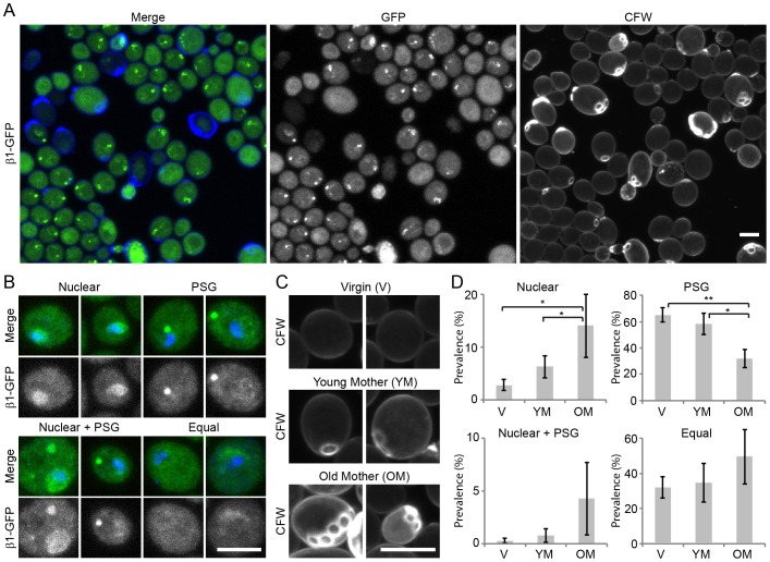
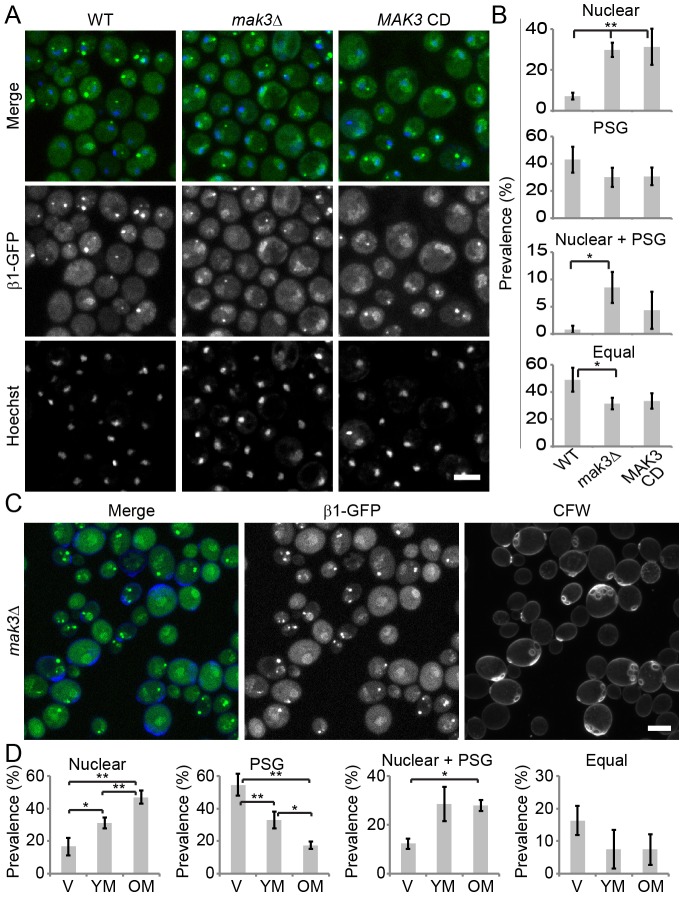
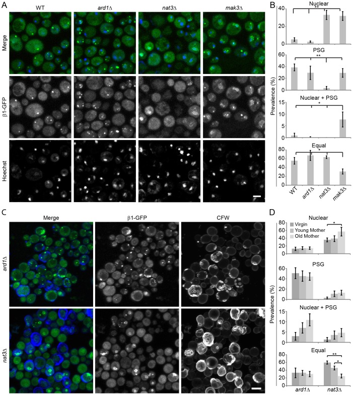
Specific degradation of proteins is essential for virtually all cellular processes and is carried out predominantly by the proteasome. The proteasome is important for clearance of damaged cellular proteins. Damaged proteins accumulate over time and excess damaged proteins can aggregate and induce the death of old cells. In yeast, the localization of the proteasome changes dramatically during aging, possibly in response to altered proteasome activity requirements. We followed two key parameters of this process: the distribution of proteasomes in nuclear and cytosolic compartments, and the formation of cytoplasmic aggregate-like structures called proteasome storage granules (PSGs). Whereas replicative young cells efficiently relocalized proteasomes from the nucleus to the cytoplasm and formed PSGs, replicative old cells were less efficient in relocalizing the proteasome and had less PSGs. By using a microscopy-based genome-wide screen, we identified genetic factors involved in these processes. Both relocalization of the proteasome and PSG formation were affected by two of the three N-acetylation complexes. These N-acetylation complexes also had different effects on the longevity of cells, indicating that each N-acetylation complex has different roles in proteasome location and aging.

Keywords: Aging; Intracellular location; N-acetylation; Proteasome; Replicative age.


Figures

Fig. 1.
Fig. 1.
Proteasome localization in nutrient-starved cells correlates with replicative age. (A) Live-cell microscopy of yeast cells in starvation shows various 20S proteasome localizations as is visualized by endogenous expression of a GFP-tagged β1 subunit (Pre3). Cells were stained with CFW to assess the replicative age of individual cells. (B) β1–GFP localization and Hoechst 33342 staining was used to define four different phenotypes: cells with cytosolic PSGs (PSG), cells with nuclear enrichment of proteasomes (Nuclear), cells that display both a nuclear enrichment of proteasomes and PSGs (Nuclear + PSG), and cells without a clear enrichment of proteasomes in PSGs or nuclei (Equal). (C) Based on CFW staining of bud scars, three different replicative age groups were defined: virgin daughter cells without bud scars (V); young mother cells with 1–2 bud scars (YM); and old mother cells with more than two bud scars (OM). (D) The prevalence of the different proteasome phenotypes in living cells from each age group was calculated by dividing the number of cells with a certain phenotype in a particular age group over the total number of cells in this age group. Results are mean±s.d. based on three independent experiments. Significance was calculated with a paired, two-tailed Student's t-test (*P<0.05, **P<0.01). Scale bars: 5 µm.
Fig. 2.
Fig. 2.
A genome-wide screen identifying genes affecting nuclear proteasome localization during starvation. (A) Schematic overview of the screening. (1) A yeast knockout library was crossed with a β1–GFP→mRFP RITE strain. (2) Tag recombination (switch) was performed after 2 days during a 5-day starvation experiment. (3) Samples were fixed, stained with Hoechst 33342 and printed on yeast arrays. (4) Microscopic imaging of GFP (old proteasomes), RFP (new proteasomes) and Hoechst 33342 (nuclei) was performed. (5) Images were analyzed by CellProfiler. (6) mak10Δ was one of the hits for a nuclear proteasome enrichment. (B) Confocal microscopy images of three hits showing nuclear enrichment of GFP-labeled proteasome in the nucleus: hul5Δ, mak10Δ and uba3Δ. Only background signal is observed for the mRFP proteasome. (C) Quantification of nuclear:cytosolic ratios of GFP in WT and nuclear retention hits. Results are mean±s.d. based on five independent experiments. Significance was calculated with a paired, two tailed Student's t-test (*P<0.05, **P<0.01). Scale bar: 5 µm.
Fig. 3.
Fig. 3.
Loss of N-acetylation by NatC causes nuclear retention of the proteasome without affecting PSG formation with both phenotypes being affected by replicative age. (A) Fixed-cell microscopy of Hoechst-33342-stained mak3Δ cells or cells expressing a catalytically inactive Mak3 (MAK3-CD) showing an increased population of cells displaying nuclear retention of proteasomes after a 5-day starvation period. (B) The prevalence of the different phenotypes in the total population was scored in three independent experiments. (C) Live-cell microscopy of mak3Δ cells stained with CFW after a 5-day starvation period. (D) The prevalence of each proteasome phenotype in living cells was scored in the three different age groups (V, virgin daughter cells; YM, young mother cells; OM, old mother cells). Results are mean±s.d. based on three independent experiments. Significance was calculated with a paired two-tailed Student's t-tests (*P<0.05, **P<0.01). Scale bars: 5 µm.
Fig. 4.
Fig. 4.
Nuclear-to-cytosolic relocalization of the proteasome during starvation requires N-acetylation by NatB and NatC. (A) Fixed-cell microscopy of starved ard1Δ (NatA deficient), nat3Δ (NatB deficient) and mak3Δ (NatC deficient) cells. Nuclei were visualized with a Hoechst 33342 staining. (B) Prevalence of the different proteasome localization phenotypes was scored in the total population. Results are mean±s.d. and are based on a biological triplicate. (C) Live-cell imaging of starved ard1Δ and nat3Δ cells. Cells were stained with CalcoFluor White to assess their replicative age. (D) Prevalence of the different proteasome phenotypes in the three age groups in living cells was quantified (mean±s.d.) in three independent experiments and significance was calculated with a paired two-tailed Student's t-test. (*P<0.05, **P<0.01). Scale bars: 5 µm.
Fig. 5.
Fig. 5.
In starvation, loss of NatA and NatB has a general effect on reproductive capacity, whereas loss of NatC specifically affects old cells. The reproductive capacity of the different strains in log phase (A) and starvation (B) was assessed by a plating assay. Loss of NatA and NatB compromised reproductive capacity in starved cells, whereas loss of NatC did not. A representative plating assay from three independent experiments is shown. (C) After CFW staining of a starved culture, the cells with the lowest (negative) and highest (positive) CFW signal were sorted to obtain populations of virgin cells and old mothers, respectively. The CFW image is the maximum projection of a 5-µm Z-stack, the DIC image is a single scan in the middle of the Z-stacks. (D) CFUs were counted for old mother (old) and virgin (young) cells. Loss of NatC affects the reproductive capacity of old cells, whereas loss of NatA and NatB reduce the reproductive capacity of both young and old cells. Results are mean±s.d. based on on three independent experiments. Significance was calculated with a paired two-tailed Student's t-test. (* = P<0.05, ** = P<0.01).

References

    1. Allen C., Büttner S., Aragon A. D., Thomas J. A., Meirelles O., Jaetao J. E., Benn D., Ruby S. W., Veenhuis M., Madeo F. et al. (2006). Isolation of quiescent and nonquiescent cells from yeast stationary-phase cultures. J. Cell Biol. 174, 89–100 10.1083/jcb.200604072 - DOI - PMC - PubMed
    1. Amm I., Sommer T., Wolf D. H. (2014). Protein quality control and elimination of protein waste: the role of the ubiquitin-proteasome system. Biochim. Biophys. Acta 1843, 182–196 10.1016/j.bbamcr.2013.06.031 - DOI - PubMed
    1. Aragon A. D., Rodriguez A. L., Meirelles O., Roy S., Davidson G. S., Tapia P. H., Allen C., Joe R., Benn D., Werner-Washburne M. (2008). Characterization of differentiated quiescent and nonquiescent cells in yeast stationary-phase cultures. Mol. Biol. Cell 19, 1271–1280 10.1091/mbc.E07-07-0666 - DOI - PMC - PubMed
    1. Arnesen T. (2011). Towards a functional understanding of protein N-terminal acetylation. PLoS Biol. 9, e1001074 10.1371/journal.pbio.1001074 - DOI - PMC - PubMed
    1. Arnesen T., Van Damme P., Polevoda B., Helsens K., Evjenth R., Colaert N., Varhaug J. E., Vandekerckhove J., Lillehaug J. R., Sherman F. et al. (2009). Proteomics analyses reveal the evolutionary conservation and divergence of N-terminal acetyltransferases from yeast and humans. Proc. Natl. Acad. Sci. USA 106, 8157–8162 10.1073/pnas.0901931106 - DOI - PMC - PubMed

Publication types

MeSH terms

Substances

LinkOut - more resources