Review
. 2015 Jun;30(3):615-32.
doi: 10.1007/s11011-014-9628-y. Epub 2014 Nov 21.

Glucose metabolism following human traumatic brain injury: methods of assessment and pathophysiological findings

Affiliations
Review

Glucose metabolism following human traumatic brain injury: methods of assessment and pathophysiological findings

Ibrahim Jalloh et al. Metab Brain Dis. 2015 Jun.

Abstract

The pathophysiology of traumatic brain (TBI) injury involves changes to glucose uptake into the brain and its subsequent metabolism. We review the methods used to study cerebral glucose metabolism with a focus on those used in clinical TBI studies. Arterio-venous measurements provide a global measure of glucose uptake into the brain. Microdialysis allows the in vivo sampling of brain extracellular fluid and is well suited to the longitudinal assessment of metabolism after TBI in the clinical setting. A recent novel development is the use of microdialysis to deliver glucose and other energy substrates labelled with carbon-13, which allows the metabolism of glucose and other substrates to be tracked. Positron emission tomography and magnetic resonance spectroscopy allow regional differences in metabolism to be assessed. We summarise the data published from these techniques and review their potential uses in the clinical setting.


Figures

Fig. 1
Fig. 1
Methods for measuring glucose metabolism in the human brain: Various techniques lend themselves to measuring glucose metabolism in the human brain. (1) Microdialysis, which allows metabolites in the extracellular fluid to be measured, is an invasive technique requiring insertion of a microdialysis catheter (possessing a semipermeable membrane, with nominal molecular weight cut-off typically 20 kDa or 100 kDa) into brain parenchyma. A pump is used to infuse perfusion fluid at a low flow rate (e.g. 0.3 μl/min) into the catheter and returned microdialysate is collected into small vials for subsequent analysis. (2) Imaging techniques include positron emission tomography (PET), which uses detectors to measure ionising radiation emitted from the glucose analogue FDG administered intravenously. This provides a measure of the uptake and phosphorylation of glucose. (3) 1H and 31P magnetic resonance spectroscopy (MRS) can be used to measure lactate and high-energy phosphate compounds respectively, in different regions of the brain. (4) A global measure of uptake or release from the brain can be achieved by using a jugular bulb catheter, which allows the venous outflow of the brain to be sampled. Measured concentrations in blood sampled from the venous catheter can be compared with arterial concentrations of metabolites, typically measured in blood taken from an arterial line. This enables the net uptake or release of metabolites by the brain to be calculated and, if cerebral blood flow is known, the cerebral metabolic rate to be calculated
Fig. 2
Fig. 2
Glycolysis and the tricarboxylic acid cycle: Glucose is the preferred substrate for the brain, although the brain can take up lactate, other monocarboxylic acids and ketone bodies under certain circumstances, for example, during the perinatal period. Once it enters cells, glucose is metabolised through glycolysis to pyruvate. Glycolysis (also termed Embden-Meyerhof pathway) is the series of reactions that results in the breakdown of glucose, generating pyruvate, adenosine triphosphate (ATP) and nicotinamide adenine dinucleotide (NADH). It is a truly fundamental pathway, found throughout nature and proceeds without the need for oxygen. The ten key enzymatic steps, in which 2 ATP molecules are consumed early on but then paid back later with the generation of 4 ATP molecules per glucose molecule, so the net production of ATP is 2 molecules per molecule of glucose, takes place in the cytoplasm. There are three key regulatory points catalysed by the enzymes hexokinase, phosphofructokinase, and pyruvate kinase. These reactions are essentially irreversible whereas the other enzymatic steps exist in equilibrium. Pyruvate is converted to acetyl CoA, which enters the tricarboxylic acid (TCA) cycle, within mitochondria. The TCA cycle results in the transfer of electrons (from NADH and succinate), to electron transport chains (ETC.) located in the inner mitochondrial membrane, which ultimately deposit on oxygen molecules. Thus the TCA cycle generates carbon dioxide (also generated by the pyruvate dehydrogenase step prior to TCA cycle) and the ETC generates water. The ETCs pump protons across the inner mitochondrial membrane, maintaining a gradient of protons across the membrane. Protons then flow down their concentration gradient, through ATP synthetases (ATPase), resulting in the generation of ATP, the cells’ widely used energy currency. Energy production from glucose is intrinsically related to neurotransmission. Glutamate spins off the TCA cycle from α-ketoglutarate (αKG), an intermediate of the TCA cycle. Glutamate can be converted reversibly into glutamine. Glutamate can also be converted into gamma-aminobutyric acid (GABA). There is a constant cycle of glutamate released during neurotransmission, retrieved from synaptic junctions by astrocytes and returned to neurons as glutamine
Fig. 3
Fig. 3
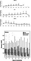
Pattern of declining microdialysate glucose after TBI: Upper panel: Graph showing initially ‘normal’ followed by declining mean microdialysate glucose concentrations after post-injury day 5 in those patients who went on to have a good outcome (Glasgow Outcome Scale extended (GOSe) 5 to 8). Figure originally published in the Journal of Cerebral Blood Flow & Metabolism (Vespa et al. 2003). Lower panel: Pooled microdialysis glucose concentrations averaged by day of monitoring and split by outcome categories demonstrating declining median concentrations over the course of a week of post-injury monitoring. Figure originally published in Brain (Timofeev et al. 2011a)
Fig. 4
Fig. 4
FDG-PET measurement of CMRglc and its relationship to brain microdialysate composition: ac FDG-PET CMRglc map demonstrating relatively high FDG uptake at sites of injury, in contrast to less injured areas of the brain. a Computed tomography (CT) scan showing gold tip of microdialysis catheter (indicated by arrow). b Co-registered FDG-PET CMRglc map showing high FDG uptake at sites of injury. c Overlay of CT and co-registered CMRglc map, showing microdialysis catheter tip location (arrow). (df Graphs illustrating relationships by linear regression (for 22 ROIs in 17 TBI patients) between FDG-PET derived CMRglc and the microdialysis parameters measured during the scan (d) lactate, (e) pyruvate, (f) lactate/pyruvate (L/P) ratio, and (g) glucose. For the linear regressions in (dg), corresponding values of p (ANOVA) are <0.0001, <0.0001, 0.74 and 0.48 respectively. Data-points from catheters at craniotomy sites (4 patients) are differentiated by grey triangles. Data-points from a second FDG-PET scan (one patient) are differentiated by grey diamonds. All other data-points are depicted as black circles (catheters inserted via cranial access device). Linear regressions presented on the graphs are for the entire (combined black plus grey symbols) dataset consisting of all 22 ROIs. Figure originally published in Acta Neurochirurgica (Hutchinson et al. 2009)
Fig. 5
Fig. 5
Carbon-13 labelling patterns after administration of 1-13C glucose: When the first carbon of glucose (C1) is 13C, the resulting pyruvate maintains the 13C as the third carbon (C3). Glycolysis converts one molecule of glucose into two molecules of pyruvate and so only one of the two 3-carbon chain length pyruvate molecules will contain 13C. Pyruvate can be converted to lactate, by the action of lactate dehydrogenase. Alternatively, through the action of pyruvate dehydrogenase on pyruvate, acetyl-CoA is produced containing just a 2-carbon backbone with one of the carbons (C1, the carboxylate carbon of pyruvate) lost as carbon dioxide. The 2 carbons of acetyl-CoA are then condensed with oxaloacetate and continue in the TCA cycle, ultimately emerging as amino acid spin-offs or as carbon dioxide. The main products identifiable with NMR (in animal brain tissue extracts ex vivo) or MRS (in human or animal brain in vivo) include glutamine, glutamate, GABA and aspartate. The C1 of a glucose molecule will become the C4 of glutamate and glutamine on the first turn of the TCA cycle. Subsequent turns of the TCA cycle will result in the 13C label shifting to the C3 or C2 of glutamate and glutamine. Hence, it is possible to determine, through the relative enrichment of the C4, C3, or C2 carbons, the proportion of glutamate or glutamine produced on the first turn of the TCA and subsequent cycles. In this way, 13C-NMR provides a convenient way to study the transformation of the carbon skeleton that takes place during the metabolism of glucose. The relative proportions of 13C incorporation at the different carbon positions of the relevant metabolites have been used in numerous studies to compare pathway activities. This schematic diagram is based on results obtained with singly labelled substrates, by Gallagher et al. (2009), Tyson et al. (2003) and Sampol et al. (2013) Abbreviations: PDH pyruvate dehydrogenase, PC, pyruvate carboxylase, ME malic enzyme
Fig. 6
Fig. 6
Illustrative 13C-NMR spectra achieved by ex vivo NMR of microdialysate after delivery of 3-13C lactate and 1,2-13C2 glucose to TBI patients: Upper panel: a and b Examples of 13C NMR spectra of brain microdialysates from a TBI patient, who received perfusion with 3-13C lactate (4 mM) by microdialysis catheters via a craniotomy (CTO); red stars indicate 13C signals for glutamine C4, C3 and C2 indicating metabolism via TCA cycle. c 13C NMR spectrum of the 3-13C lactate substrate solution prior to perfusing. d 13C NMR spectrum of brain microdialysate from an unlabelled patient. For further details, see Gallagher et al. (2009). Figure originally published in Brain (Gallagher et al. 2009). Lower panel: Examples of 13C NMR spectra of brain microdialysates from patients who received 1,2-13C2 glucose (4 mM) perfused via the microdialysis catheter. Uninjured brain is normal-appearing brain in a patient operated on for a benign tumour elsewhere in the brain. TBI brain is from a patient with a diffuse injury. The part of the spectrum illustrated in each case is for the C3 carbon of lactate. Also present in this part of the spectrum is one of the signals due to the internal standard DSS (2,2-dimethyl-2-silapentane-5-sulfonate sodium salt). The remainder of the spectrum (including the main DSS signal at 0 ppm) is not shown. The C3 doublet indicated by red stars represents lactate doubly labelled with 13C, produced by glycolysis; the C3 signal for 13C is split into 2 peaks by coupling to 13C also present at the neighbouring C2 position within the same molecule. The C3 singlet indicated by green stars represents lactate singly labelled with 13C, produced via the PPP. Figure originally published in European Journal of Pharmaceutical Sciences (Carpenter et al. 2014)

Similar articles

Cited by

References

    1. Aaen GS, Holshouser BA, Sheridan C, et al. Magnetic resonance spectroscopy predicts outcomes for children with nonaccidental trauma. Pediatrics. 2010;125:295–303. doi: 10.1542/peds.2008-3312. - DOI - PubMed
    1. Alessandri B, Rice AC, Levasseur J, et al. Cyclosporin A improves brain tissue oxygen consumption and learning/memory performance after lateral fluid percussion injury in rats. J Neurotrauma. 2002;19:829–841. doi: 10.1089/08977150260190429. - DOI - PubMed
    1. Andersen BJ, Marmarou A. Post-traumatic selective stimulation of glycolysis. Brain Res. 1992;585:184–189. doi: 10.1016/0006-8993(92)91205-S. - DOI - PubMed
    1. Ashwal S, Holshouser BA, Shu SK, et al. Predictive value of proton magnetic resonance spectroscopy in pediatric closed head injury. Pediatr Neurol. 2000;23:114–125. doi: 10.1016/S0887-8994(00)00176-4. - DOI - PubMed
    1. Bartnik BL, Lee SM, Hovda DA, Sutton RL. The fate of glucose during the period of decreased metabolism after fluid percussion injury: a 13C NMR study. J Neurotrauma. 2007;24:1079–1092. doi: 10.1089/neu.2006.0210. - DOI - PubMed

Publication types

LinkOut - more resources