. 2015 Mar;235(4):593-605.
doi: 10.1002/path.4486. Epub 2014 Dec 17.

Characterization of the cell of origin and propagation potential of the fibroblast growth factor 9-induced mouse model of lung adenocarcinoma

Affiliations

Characterization of the cell of origin and propagation potential of the fibroblast growth factor 9-induced mouse model of lung adenocarcinoma

Daisuke Arai et al. J Pathol. 2015 Mar.

Abstract

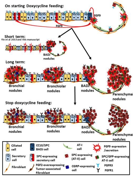
Fibroblast growth factor 9 (FGF9) is essential for lung development and is highly expressed in a subset of human lung adenocarcinomas. We recently described a mouse model in which FGF9 expression in the lung epithelium caused proliferation of the airway epithelium at the terminal bronchioles and led to rapid development of adenocarcinoma. Here, we used this model to characterize the effects of prolonged FGF9 induction on the proximal and distal lung epithelia, and examined the propagation potential of FGF9-induced lung tumours. We showed that prolonged FGF9 over-expression in the lung resulted in the development of adenocarcinomas arising from both alveolar type II and airway secretory cells in the lung parenchyma and airways, respectively. We found that tumour cells harboured tumour-propagating cells that were able to form secondary tumours in recipient mice, regardless of FGF9 expression. However, the highest degree of tumour propagation was observed when unfractionated tumour cells were co-administered with autologous, tumour-associated mesenchymal cells. Although the initiation of lung adenocarcinomas was dependent on activation of the FGF9-FGF receptor 3 (FGFR3) signalling axis, maintenance and propagation of the tumour was independent of this signalling. Activation of an alternative FGF-FGFR axis and the interaction with tumour stromal cells is likely to be responsible for the development of this independence. This study demonstrates the complex role of FGF-FGFR signalling in the initiation, growth and propagation of lung cancer. Our findings suggest that analysing the expressions of FGF-FGFRs in human lung cancer will be a useful tool for guiding customized therapy.

Keywords: FGF9; FGFR; adenocarcinoma; lung cancer; tumour propagation; tumour-associated lung fibroblasts.


Figures

Figure 1
Figure 1. The effects of prolonged FGF9 overexpression on the lungs
DT mice were induced with doxycycline for 6 months. Representative lung sections show (A) the presence of tumor nodules in bronchi, in mid-level bronchioles, near BADJ and in the lung parenchyma. (B) FGF9 overexpression in the lung caused many of the epithelial cells lining the airways (primarily Club cells) to upregulate Sftpc expression and thus activated the FGF9/GFP transgene in them. (CE) Examination of the lungs of wt mice 2-days after intratracheal administration of recombinant FGF9 revealed that many Club cells in the proximal bronchi, in mid-level bronchioles, and near the BADJ expressed Sftpc. (F) Both airways and lung parenchymal nodules expressed the pan-epithelial marker E-cadherin. (G) Phenotypic heterogeneity, with many GFP negative cells are observed within each nodule along with PCNA-positive (proliferating) cells that were both GFP-positive and negative. Scale bars: (A) 200 μm; (B, F, G) 100 μm; (C–E) 50 μm. See also Supplementary Figures S1–S3.
Figure 2
Figure 2. Propagation potential of the FGF9-induced adenocarcinoma
(A) Schematic diagram summarizing the design and main findings of the tumor propagation study. (B–E) MicroCT is a sensitive tool for the early detection of tumor shadows in mouse lungs. CT images from wt syngeneic (n=3) (B) and all DT mice before the start of the doxycycline feeding (C) showed nodule-free lungs. (D) Four weeks after the start of doxycycline feeding, the lungs of the DT mice that were intended for use as a cell source for the tumor propagation experiment exhibited several tumor shadows (red arrows, n=20). (E) A wt syngeneic mouse that was subcutaneously injected with WLCs from the doxycycline-fed DT mice had a large nodular shadow in its lung 1-month after the subcutaneous injection (red arrow, n=1). (F) Secondary tumors stained with H&E showed histological characteristics that recapitulated those of primary lung adenocarcinoma. (G-H) Immunofluorescent staining of the secondary tumor nodules confirmed its similarity to primary lung parenchymal nodules (expressions of Sftpc and AQP5, but not that of CC10 or CGRP). (I) Lungs of recipient mice had accumulation of inflammatory cells around blood vessels and bronchi/bronchioles,; BALT and perivascular cuffing. (J-K) BALT and perivascular cuffs stained positive for the pan-hematopoietic marker CD45 and the T-lymphocytic marker CD3, confirming their identity. Scale bars: (D) 200 μm; (E–I) 100 μm. See also Supplementary Figure S4.
Figure 3
Figure 3. TALF-induced colony/sphere formation by epithelial cells possibly through FGF2/FGFR signaling
Fibroblasts isolated from the lungs of a wt (A) or tumor-bearing DT mice lungs (i.e.TALF) (B) were co-cultured with wt lung epithelium in the 3D-sphere/colony formation organoid assay and were observed for 7-days (n=3, independent experiments). TALF co-culture produced epithelial spheres that were of larger size and number than the spheres observed in the co-culture with wt fibroblasts. (C) Quantitative real-time PCR analysis comparing the gene expressions of FGF2, FGF9, FGF10, FGFR1, FGFR2, and FGFR3 between TALFs, wt fibroblasts, and a mouse lung epithelial cell line. FGF2 and FGFR2 expression levels were significantly higher in TALFs than in the wt fibroblasts (* p <0.001). (D) Wt lung epithelial cells were co-cultured with TALFs and treated with an FGFR inhibitor. Blocking of FGFRs resulted in formation of smaller and fewer colonies. (E) Quantification of sphere number and diameter. The results confirmed that TALFs significantly induced clonal growth of epithelial progenitors cells compared to wt fibroblasts. (*,# p<0.01 compared to A and B, respectively).
Figure 4
Figure 4. Effect of FGF9 withdrawal on established lung tumors
DT mice were fed doxycycline chow for 1-month (n=4), and the presence of tumor nodules in their lungs was confirmed by microCT (A, D and G). Then, they were switched into dox-free chow, and the status of tumor nodules in their lungs was examined monthly. Many of the previously identified nodules persisted, and their size remained the same or increased (compare B and C to A, and E and F to D). Some nodules appeared de novo after doxycycline withdrawal and increased in size between months 3 and 4 (compare H and I to G). The role of signaling through the FGF/FGFR pathway. (J-K) EpCAM+ epithelial cells were sorted from dox-fed, DT mouse lungs and then co-cultured with wt fibroblasts in the 3D-sphere/colony formation assay. Culture wells were treated with the FGFR inhibitor or a vehicle control (n=3 well replicates, two independent experiments). FGFR inhibitor-treated cells formed spheres that were of smaller size and fewer number than those in the control group. (L) Quantification of sphere number and diameter. (*p<0.01, compared to J). Scale bar: (J) 100 μm. See also Supplementary Figures S5–S7.
Figure 5
Figure 5. Summary of the response of various respiratory cells to FGF9 induction and withdrawal
On feeding doxycycline to the Sftpc-rtTA and Tre-Fgf9-ires-eGfp double-transgenic mice, Sftpc-expressing cells started to express FGF9 and GFP. Secreted FGF9 binds to its receptor, FGFR3, which is constitutively expressed in all lung epithelial cells. Within hours to days, adenomas and adenocarcinomas develop, mostly as a result of proliferation of epithelial cells at BADJ (15). Furthermore, FGF9 induces airway secretory cells to overexpress Sftpc and thus the FGF9/GFP transgene is switched in these cells. Within weeks to months, tumor nodules develop in bronchi, bronchioles, and lung parenchyma. FGFR1 and FGF2 expressions were upregulated in tumor epithelial cells and lung fibroblasts, respectively. FGFR3 expression gradually diminishes. Tumor nodules contained both GFP-positive and GFP-negative cells, and bronchial nodules expressed CC10 and CGRP. After withdrawal of doxycycline feeding for 3 months, FGF9/GFP expression was lost, but tumor nodules persisted or continued to grow. Tumor epithelial cells continued to express FGFR1.

References

    1. Imielinski M, Berger AH, Hammerman PS, et al. Mapping the hallmarks of lung adenocarcinoma with massively parallel sequencing. Cell. 2012;150:1107–1120. - PMC - PubMed
    1. Ding L, Getz G, Wheeler DA, et al. Somatic mutations affect key pathways in lung adenocarcinoma. Nature. 2008;455:1069–1075. - PMC - PubMed
    1. Taron M, Ichinose Y, Rosell R, et al. Activating mutations in the tyrosine kinase domain of the epidermal growth factor receptor are associated with improved survival in gefitinib-treated chemorefractory lung adenocarcinomas. Clin Cancer Res. 2005;11:5878–5885. - PubMed
    1. Nguyen KS, Kobayashi S, Costa DB. Acquired resistance to epidermal growth factor receptor tyrosine kinase inhibitors in non-small-cell lung cancers dependent on the epidermal growth factor receptor pathway. Clin Lung Cancer. 2009;10:281–289. - PMC - PubMed
    1. Boch C, Kollmeier J, Roth A, et al. The frequency of EGFR and KRAS mutations in non-small cell lung cancer (NSCLC): routine screening data for central Europe from a cohort study. BMJ Open. 2013;3:e002560. - PMC - PubMed

Publication types

MeSH terms

Substances