

The type I BMP receptor ACVR1/ALK2 is required for chondrogenesis during development
- PMID: 25413979
- PMCID: PMC4376569
- DOI: 10.1002/jbmr.2385
The type I BMP receptor ACVR1/ALK2 is required for chondrogenesis during development
Abstract
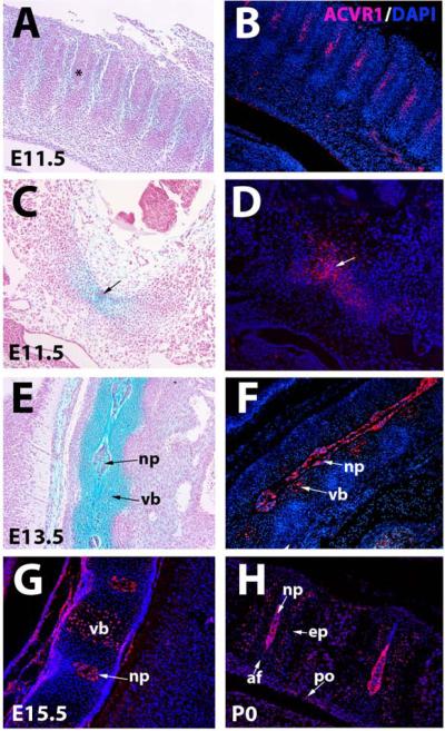
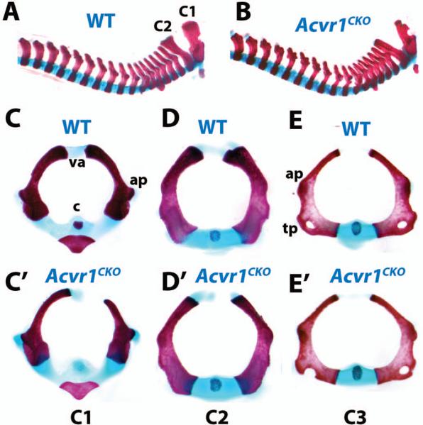
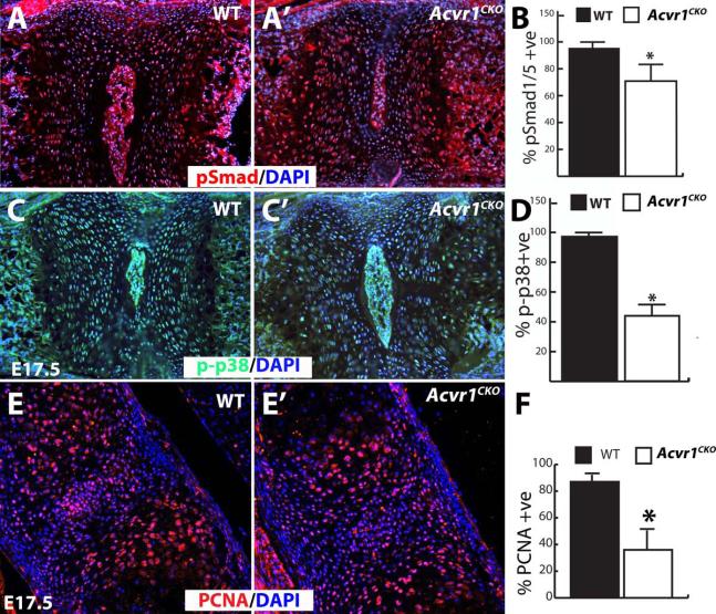
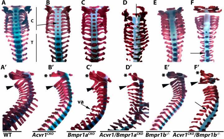
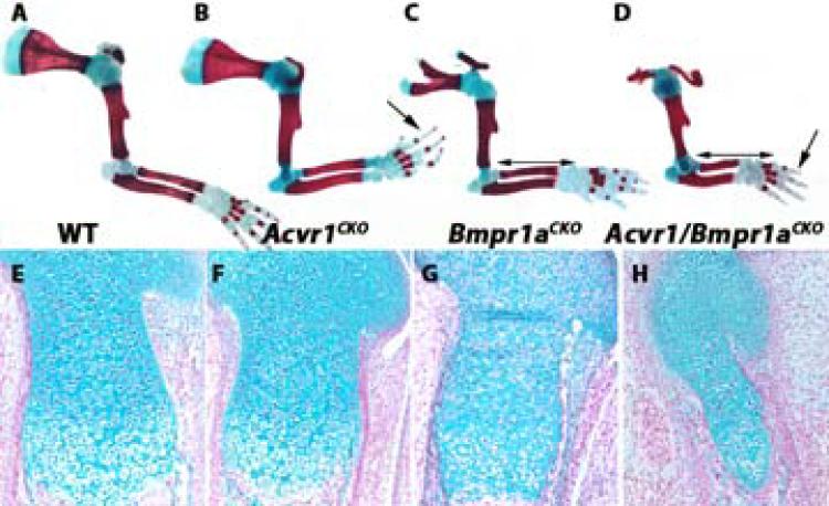
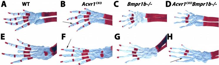
Bone morphogenetic proteins (BMPs) are crucial regulators of chondrogenesis. BMPs transduce their signals through three type I receptors: BMPR1A, BMPR1B, and ACVR1/ALK2. Fibrodysplasia ossificans progressiva (FOP), a rare disorder characterized by progressive ossification of connective tissue, is caused by an activating mutation in Acvr1 (the gene that encodes ACVR1/ALK2). However, there are few developmental defects associated with FOP. Thus, the role of ACVR1 in chondrogenesis during development is unknown. Here we report the phenotype of mice lacking ACVR1 in cartilage. Acvr1(CKO) mice are viable but exhibit defects in the development of cranial and axial structures. Mutants exhibit a shortened cranial base, and cervical vertebrae are hypoplastic. Acvr1(CKO) adult mice develop progressive kyphosis. These morphological defects were associated with decreased levels of Smad1/5 and p38 activation, and with reduced rates of chondrocyte proliferation in vertebral cartilage. We also tested whether ACVR1 exerts coordinated functions with BMPR1A and BMPR1B through analysis of double mutants. Acvr1/Bmpr1a and Acvr1/Bmpr1b mutant mice exhibited generalized perinatal lethal chondrodysplasia that was much more severe than in any of the corresponding mutant strains. These findings demonstrate that ACVR1 is required for chondrocyte proliferation and differentiation, particularly in craniofacial and axial elements, but exerts coordinated functions with both BMPR1A and BMPR1B throughout the developing endochondral skeleton.
Keywords: ACVR1; ALK2; BMP; CHONDROGENESIS; MOUSE.
© 2014 American Society for Bone and Mineral Research.
Figures
Similar articles
-
Dysregulated BMP signaling through ACVR1 impairs digit joint development in fibrodysplasia ossificans progressiva (FOP).Dev Biol. 2021 Feb;470:136-146. doi: 10.1016/j.ydbio.2020.11.004. Epub 2020 Nov 17. Dev Biol. 2021. PMID: 33217406 Free PMC article.
-
ACVR1-Fc suppresses BMP signaling and chondro-osseous differentiation in an in vitro model of Fibrodysplasia ossificans progressiva.Bone. 2016 Nov;92:29-36. doi: 10.1016/j.bone.2016.07.023. Epub 2016 Aug 2. Bone. 2016. PMID: 27492611
-
Neofunction of ACVR1 in fibrodysplasia ossificans progressiva.Proc Natl Acad Sci U S A. 2015 Dec 15;112(50):15438-43. doi: 10.1073/pnas.1510540112. Epub 2015 Nov 30. Proc Natl Acad Sci U S A. 2015. PMID: 26621707 Free PMC article.
-
Common mutations in ALK2/ACVR1, a multi-faceted receptor, have roles in distinct pediatric musculoskeletal and neural orphan disorders.Cytokine Growth Factor Rev. 2016 Feb;27:93-104. doi: 10.1016/j.cytogfr.2015.12.007. Epub 2015 Dec 28. Cytokine Growth Factor Rev. 2016. PMID: 26776312 Free PMC article. Review.
-
Structural basis for the potent and selective binding of LDN-212854 to the BMP receptor kinase ALK2.Bone. 2018 Apr;109:251-258. doi: 10.1016/j.bone.2017.09.004. Epub 2017 Sep 12. Bone. 2018. PMID: 28918311 Free PMC article. Review.
Cited by
-
Bone morphogenetic protein receptor signal transduction in human disease.J Pathol. 2019 Jan;247(1):9-20. doi: 10.1002/path.5170. Epub 2018 Nov 27. J Pathol. 2019. PMID: 30246251 Free PMC article. Review.
-
The TGFβ type I receptor TGFβRI functions as an inhibitor of BMP signaling in cartilage.Proc Natl Acad Sci U S A. 2019 Jul 30;116(31):15570-15579. doi: 10.1073/pnas.1902927116. Epub 2019 Jul 16. Proc Natl Acad Sci U S A. 2019. PMID: 31311865 Free PMC article.
-
TGF-β Family Signaling in Connective Tissue and Skeletal Diseases.Cold Spring Harb Perspect Biol. 2017 Nov 1;9(11):a022269. doi: 10.1101/cshperspect.a022269. Cold Spring Harb Perspect Biol. 2017. PMID: 28246187 Free PMC article. Review.
-
The transcriptional cofactor Jab1/Cops5 is crucial for BMP-mediated mouse chondrocyte differentiation by repressing p53 activity.J Cell Physiol. 2021 Aug;236(8):5686-5697. doi: 10.1002/jcp.30254. Epub 2021 Jan 3. J Cell Physiol. 2021. PMID: 33393086 Free PMC article.
-
Palovarotene Inhibits Heterotopic Ossification and Maintains Limb Mobility and Growth in Mice With the Human ACVR1(R206H) Fibrodysplasia Ossificans Progressiva (FOP) Mutation.J Bone Miner Res. 2016 Sep;31(9):1666-75. doi: 10.1002/jbmr.2820. Epub 2016 Mar 12. J Bone Miner Res. 2016. PMID: 26896819 Free PMC article.
References
-
- Chen D, Zhao M, Mundy GR. Bone morphogenetic proteins. Growth Factors. 2004;22(4):233–41. - PubMed
-
- Clarke TR, Hoshiya Y, Yi SE, Liu X, Lyons KM, Donahoe PK. Mullerian inhibiting substance signaling uses a bone morphogenetic protein (BMP)-like pathway mediated by ALK2 and induces SMAD6 expression. Mol Endocrinol. 2001;15(6):946–59. - PubMed
-
- Luo J, Tang M, Huang J, He BC, Gao JL, Chen L, Zuo GW, Zhang W, Luo Q, Shi Q, Zhang BQ, Bi Y, Luo X, Jiang W, Su Y, Shen J, Kim SH, Huang E, Gao Y, Zhou JZ, Yang K, Luu HH, Pan X, Haydon RC, Deng ZL, He TC. TGFbeta/BMP type I receptors ALK1 and ALK2 are essential for BMP9-induced osteogenic signaling in mesenchymal stem cells. J Biol Chem. 2010;285(38):29588–98. - PMC - PubMed
-
- Haas AR, Tuan RS. Chondrogenic differentiation of murine C3H10T1/2 multipotential mesenchymal cells: II. Stimulation by bone morphogenetic protein-2 requires modulation of N-cadherin expression and function. Differentiation. 1999;64(2):77–89. - PubMed
Publication types
MeSH terms
Substances
Grants and funding
LinkOut - more resources
Full Text Sources
Other Literature Sources
Molecular Biology Databases
