. 2015 Jan 2;290(1):384-95.
doi: 10.1074/jbc.M114.590158. Epub 2014 Nov 20.

MicroRNA-340-mediated degradation of microphthalmia-associated transcription factor (MITF) mRNA is inhibited by coding region determinant-binding protein (CRD-BP)

Affiliations

MicroRNA-340-mediated degradation of microphthalmia-associated transcription factor (MITF) mRNA is inhibited by coding region determinant-binding protein (CRD-BP)

Srikanta Goswami et al. J Biol Chem. .

Abstract

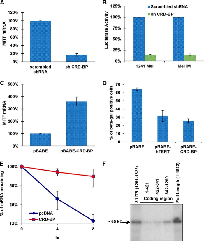
Alternative cleavage and polyadenylation generates multiple transcript variants producing mRNA isoforms with different length 3'-UTRs. Alternative cleavage and polyadenylation enables differential post-transcriptional regulation via the availability of different cis-acting elements in 3'-UTRs. Microphthalmia-associated transcription factor (MITF) is a master regulator of melanocyte development and melanogenesis. This central transcription factor is also implicated in melanoma development. Here, we show that melanoma cells favor the expression of MITF mRNA with a shorter 3'-UTR. We also establish that this isoform is regulated by a micro RNA (miRNA/miR), miR-340. miR-340 interacts with two of its target sites on the MITF 3'-UTR, causing mRNA degradation as well as decreased expression and activity of MITF. Conversely, the RNA-binding protein, coding region determinant-binding protein, was shown to be highly expressed in melanoma, directly binds to the 3'-UTR of MITF mRNA, and prevents the binding of miR-340 to its target sites, resulting in the stabilization of MITF transcripts, elevated expression, and transcriptional activity of MITF. This regulatory interplay between RNA-binding protein and miRNA highlights an important mechanism for the regulation of MITF in melanocytes and malignant melanomas.

Keywords: Alternative Cleavage and Polyadenylation; CRD-BP; IGF2BP1; MITF; Melanogenesis; Melanoma; MicroRNA (miRNA); Ribonuclear Protein (RNP); mRNA; mRNA Decay; miRNA.


Figures

FIGURE 1.
FIGURE 1.
MITF mRNA with a short 3′-UTR is more abundant in melanoma cell lines. A, graphical representation of different MITF mRNA isoforms with varying 3′-UTR lengths. The approximate positions of reported miRNA target sites are shown on long and medium 3′-UTRs. B, expression of MITF mRNA isoforms containing various 3′-UTR lengths evaluated by qRT-PCR using primers specific for the long, medium, and short 3′-UTR.
FIGURE 2.
FIGURE 2.
The abundant short 3′-UTR MITF mRNA is also regulated by miRNA. A, Dicerwt and DicerEx5/Ex5 HCT116 cells were co-transfected with Tet-Off plasmid and p-BI-G-MITF plasmid with a short 3′-UTR. Transcription was stopped by treatment with doxycycline for the indicated durations. The stability of MITF transcripts was analyzed by measuring MITF mRNA levels with qRT-PCR and normalized to GAPDH expression. B, p-BI-G-MITF plasmid with short 3′-UTR was expressed in DLD1 cells, Dicerwt and DicerEx5/Ex5, under the control of the Tet-Off system. The stability of MITF mRNA was analyzed as in A. C, the stability of MITF transcripts with short 3′-UTR expressed in RKOwt and RKO DicerEx5/Ex5 cells was analyzed as in A. All results are representative of three separate experiments and expressed as the mean ± S.D. (error bars). The average half-lives of mRNAs are presented in Table 3. D, sequence of the short 3′-UTR of MITF showing binding sites for miR-340 (in boldface type) and miR-548c-3p (underlined). E, the levels of endogenous MITF mRNA in 451Lu cells, transfected with the indicated miR-sponge constructs, were estimated by qRT-PCR after normalization to GAPDH expression. Results are representative of three separate experiments and presented as a percentage of control (SP-CXCR4) as the mean ± S.D. (error bars). F, immunoblot analysis of MITF expression in 451Lu cells transfected with indicated miR-sponge constructs (top panel). The lower panel shows β-actin expression.
FIGURE 3.
FIGURE 3.
MITF mRNA is a target of miR-340. A, 451Lu cells were co-transfected with Tet-Off plasmid, p-BI-G-MITF plasmid with short 3′-UTR, and the indicated miR-sponge construct. Transcription was stopped by treatment with doxycycline for the indicated durations. The stability of MITF transcripts was analyzed by measuring MITF mRNA levels with qRT-PCR, normalized to GAPDH expression. B, 451Lu cells were co-transfected with Tet-Off plasmid, p-BI-GL-MITF-Short 3′-UTR, and the indicated miR-sponge construct. The turnover of chimeric luciferase-MITF-3′-UTR transcripts was analyzed as in A. C, 451Lu cells were co-transfected with β-gal-expressing plasmid, Tet-Off plasmid, p-BI-GL-MITFwt, or p-BI-GL-MITFδmiR-340 and the indicated miR-sponge construct. After 24 h, luciferase activity was measured. Values represent luciferase activity normalized to β-gal. D, 451Lu cells were co-transfected with Tet-Off plasmid, p-BI-G-MITFwt, or p-BI-G-MITFδmiR-340. The stability of MITF transcripts was analyzed as in A. All results are representative of three separate experiments and are expressed as the mean ± S.D. (error bars). The average half-lives of mRNAs are presented in Table 3.
FIGURE 4.
FIGURE 4.
CRD-BP is a positive regulator of MITF expression. A, 451Lu cells were transfected with either control shRNA or shRNA against CRD-BP. 48 h after transfection, cells were collected and assayed for levels of MITF mRNA by qRT-PCR, normalized to GAPDH expression, and presented as a percentage of control (scrambled shRNA). B, 1241 mel and mel IM cells were co-transfected with the indicated shRNA-expressing plasmids, β-gal-expressing plasmid, and pHTRPL4, where the luciferase gene is expressed under an MITF-dependent promoter. Values represent luciferase activity normalized to β-gal expression. C, NHMs were electroporated using the AMAXA NucleofectorTM with either CRD-BP-overexpressing plasmid or empty vector. 72 h after transfection, cells were collected and assayed for levels of MITF mRNA by qRT-PCR, normalized to GAPDH expression, and presented in percentage to control (pBABE). D, NHMs were electroporated using the AMAXA NucleofectorTM with the indicated plasmids. 48 h after electroporation, cells were stained for senescence-associated β-gal, and the percentage of β-gal-positive cells was calculated. E, 451Lu cells were co-transfected with Tet-Off plasmid, p-BI-G-MITF, and either pcDNA control or CRD-BP-overexpressing plasmid. The turnover of MITF transcripts was analyzed by qRT-PCR after stopping transcription by doxycycline treatment for the indicated times. Normalization was done with respect to GAPDH expression. All results are representative of three separate experiments and expressed as the mean ± S.D. (error bars). The average half-lives of mRNAs are presented in Table 3. F, FLAG immunoprecipitation of UV-cross-linked RNP complexes. Protein extracts from 293T cells transfected with FLAG-CRD-BP were incubated with internally 32P-labeled RNA from three fragments of the MITF mRNA coding region, the full-length MITF mRNA, and the short 3′-UTR. Fragment 1 contains nucleotides 1–421, fragment 2 consists of nucleotides 422–841, and fragment 3 consists of nucleotides 842–1260 of the MITF coding region. RNP complexes were precipitated with anti-FLAG antibodies and analyzed on PAGE and autoradiographed.
FIGURE 5.
FIGURE 5.
CRD-BP counteracts miR-340 action and stabilizes MITF mRNA. A, wild type MITF expressing plasmid p-BI-G-MITFwt was co-transfected with Tet-Off plasmid and the indicated constructs in 451Lu cells. The turnover of MITF transcripts was analyzed as in Fig. 4E. B, 451Lu cells were transfected with the indicated constructs. 48 h after transfection, cells were collected and assayed for levels of endogenous MITF mRNA by qRT-PCR, normalized to GAPDH expression, and presented as a percentage of control. C, 451Lu cells were transfected with either control pcDNA 3.1, CRD-BP-overexpressing, control pCMV-MIR, or miR-340 overexpressing (pCMV-MIR340) plasmids, as indicated. 48 h after transfection, cells were collected and assayed for levels of MITF mRNA by qRT-PCR, normalized to GAPDH expression, and presented as a percentage of control. D, 451Lu cells were co-transfected with MITF-dependent luciferase expressing vector pHTRPL4, indicated shRNA, miR-sponge-expressing plasmids, and β-gal-expressing plasmid. Values correspond to luciferase activity normalized to β-gal expression. E, construct expressing MITF with both of the miR-340 sites deleted (p-BI-G-MITFδmiR-340) was co-transfected with Tet-Off plasmid and the indicated constructs in 451Lu cells. The turnover of MITF transcript was analyzed as in Fig. 4E. All results are representative of three separate experiments and expressed as the mean ± S.D. (error bars). The average half-lives of mRNAs are presented in Table 3. F, 451Lu cells were transfected as indicated. 48 h after transfection, cells were collected and assayed for levels of mature miR-340 transcript by ddPCR. Results are presented as the number of copies/1 μg of total RNA. G, 451Lu cells were transfected as indicated. 48 h after transfection, cells were collected and assayed for levels of CRD-BP mRNA by qRT-PCR, normalized to GAPDH expression, and presented as -fold change compared with control. H, 451Lu and 928 mel cells were co-transfected with the indicated constructs and pTk-Puro. The colonies were selected for puromycin resistance, stained with crystal violet, and counted.
FIGURE 5.
FIGURE 5.
CRD-BP counteracts miR-340 action and stabilizes MITF mRNA. A, wild type MITF expressing plasmid p-BI-G-MITFwt was co-transfected with Tet-Off plasmid and the indicated constructs in 451Lu cells. The turnover of MITF transcripts was analyzed as in Fig. 4E. B, 451Lu cells were transfected with the indicated constructs. 48 h after transfection, cells were collected and assayed for levels of endogenous MITF mRNA by qRT-PCR, normalized to GAPDH expression, and presented as a percentage of control. C, 451Lu cells were transfected with either control pcDNA 3.1, CRD-BP-overexpressing, control pCMV-MIR, or miR-340 overexpressing (pCMV-MIR340) plasmids, as indicated. 48 h after transfection, cells were collected and assayed for levels of MITF mRNA by qRT-PCR, normalized to GAPDH expression, and presented as a percentage of control. D, 451Lu cells were co-transfected with MITF-dependent luciferase expressing vector pHTRPL4, indicated shRNA, miR-sponge-expressing plasmids, and β-gal-expressing plasmid. Values correspond to luciferase activity normalized to β-gal expression. E, construct expressing MITF with both of the miR-340 sites deleted (p-BI-G-MITFδmiR-340) was co-transfected with Tet-Off plasmid and the indicated constructs in 451Lu cells. The turnover of MITF transcript was analyzed as in Fig. 4E. All results are representative of three separate experiments and expressed as the mean ± S.D. (error bars). The average half-lives of mRNAs are presented in Table 3. F, 451Lu cells were transfected as indicated. 48 h after transfection, cells were collected and assayed for levels of mature miR-340 transcript by ddPCR. Results are presented as the number of copies/1 μg of total RNA. G, 451Lu cells were transfected as indicated. 48 h after transfection, cells were collected and assayed for levels of CRD-BP mRNA by qRT-PCR, normalized to GAPDH expression, and presented as -fold change compared with control. H, 451Lu and 928 mel cells were co-transfected with the indicated constructs and pTk-Puro. The colonies were selected for puromycin resistance, stained with crystal violet, and counted.
FIGURE 6.
FIGURE 6.
Model for interference of miR-340 function by CRD-BP. A, in the absence of CRD-BP, the miR-340-guided miRNA-induced silencing complex (miRISC) interacts with the 3′-UTR of MITF mRNA and accelerates its degradation. B, when CRD-BP is present, it binds to the 3′-UTR of MITF mRNA in the vicinity of miR-340 binding sites, shielding the MITF mRNA from miR-340-mediated down-regulation. The resulting elevated levels of MITF contribute to melanoma cell proliferation.

Similar articles

Cited by

References

    1. Hallsson J. H., Haflidadóttir B. S., Stivers C., Odenwald W., Arnheiter H., Pignoni F., Steingrímsson E. (2004) The basic helix-loop-helix leucine zipper transcription factor Mitf is conserved in Drosophila and functions in eye development. Genetics 167, 233–241 - PMC - PubMed
    1. Bentley N. J., Eisen T., Goding C. R. (1994) Melanocyte-specific expression of the human tyrosinase promoter: activation by the microphthalmia gene product and role of the initiator. Mol. Cell. Biol. 14, 7996–8006 - PMC - PubMed
    1. Hemesath T. J., Steingrímsson E., McGill G., Hansen M. J., Vaught J., Hodgkinson C. A., Arnheiter H., Copeland N. G., Jenkins N. A., Fisher D. E. (1994) Microphthalmia, a critical factor in melanocyte development, defines a discrete transcription factor family. Genes Dev. 8, 2770–2780 - PubMed
    1. Yasumoto K., Yokoyama K., Shibata K., Tomita Y., Shibahara S. (1994) Microphthalmia-associated transcription factor as a regulator for melanocyte-specific transcription of the human tyrosinase gene. Mol. Cell. Biol. 14, 8058–8070 - PMC - PubMed
    1. Carreira S., Liu B., Goding C. R. (2000) The gene encoding the T-box factor Tbx2 is a target for the microphthalmia-associated transcription factor in melanocytes. J. Biol. Chem. 275, 21920–21927 - PubMed

Publication types

MeSH terms

LinkOut - more resources