Review
. 2014 Nov 4:5:429.
doi: 10.3389/fphys.2014.00429. eCollection 2014.

Circadian rhythms, Wnt/beta-catenin pathway and PPAR alpha/gamma profiles in diseases with primary or secondary cardiac dysfunction

Affiliations
Review

Circadian rhythms, Wnt/beta-catenin pathway and PPAR alpha/gamma profiles in diseases with primary or secondary cardiac dysfunction

Yves Lecarpentier et al. Front Physiol. .

Abstract

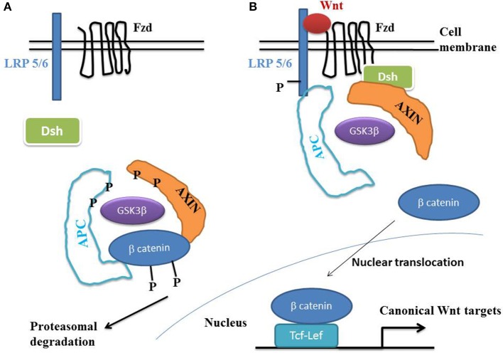
Circadian clock mechanisms are far-from-equilibrium dissipative structures. Peroxisome proliferator-activated receptors (PPAR alpha, beta/delta, and gamma) play a key role in metabolic regulatory processes, particularly in heart muscle. Links between circadian rhythms (CRs) and PPARs have been established. Mammalian CRs involve at least two critical transcription factors, CLOCK and BMAL1 (Gekakis et al., 1998; Hogenesch et al., 1998). PPAR gamma plays a major role in both glucose and lipid metabolisms and presents circadian properties which coordinate the interplay between metabolism and CRs. PPAR gamma is a major component of the vascular clock. Vascular PPAR gamma is a peripheral regulator of cardiovascular rhythms controlling circadian variations in blood pressure and heart rate through BMAL1. We focused our review on diseases with abnormalities of CRs and with primary or secondary cardiac dysfunction. Moreover, these diseases presented changes in the Wnt/beta-catenin pathway and PPARs, according to two opposed profiles. Profile 1 was defined as follows: inactivation of the Wnt/beta-catenin pathway with increased expression of PPAR gamma. Profile 2 was defined as follows: activation of the Wnt/beta-catenin pathway with decreased expression of PPAR gamma. A typical profile 1 disease is arrhythmogenic right ventricular cardiomyopathy, a genetic cardiac disease which presents mutations of the desmosomal proteins and is mainly characterized by fatty acid accumulation in adult cardiomyocytes mainly in the right ventricle. The link between PPAR gamma dysfunction and desmosomal genetic mutations occurs via inactivation of the Wnt/beta-catenin pathway presenting oscillatory properties. A typical profile 2 disease is type 2 diabetes, with activation of the Wnt/beta-catenin pathway and decreased expression of PPAR gamma. CRs abnormalities are present in numerous pathologies such as cardiovascular diseases, sympathetic/parasympathetic dysfunction, hypertension, diabetes, neurodegenerative diseases, cancer which are often closely inter-related.

Keywords: PPAR; Wnt/beta-catenin; circadian rhythms; colon cancer; diabetes; hypertension; myocardial ischemia; neurodegenerative diseases.


Figures

Figure 1
Figure 1
The Wnt/beta-catenin pathway. (A) In the absence of Wnt, cytosolic beta-catenin is phosphorylated by GSK3 beta. APS and AXIN complex with GSK3 beta and beta-catenin to enhance the destruction process into the proteasome. Phosphorylated beta-catenin is recognized by the ubiquitin ligase beta -TrCP, ubiquinated and degraded. The Wnt pathway is in an “off state.” (B) In the presence of Wnt, Wnt binds both Frizzled and LRP5/6 receptors to initiate GRK5/6-mediated LRP phosphorylation and disheveled-mediated Frizzled internalization. Disheveled membrane translocation leads to dissociation of the AXIN/APC/GSK3 beta complex. Beta-catenin phosphorylation is inhibited and accumulates into the cytosol. Beta-catenin then translocates to the nucleus to bind Lef-Tcf co-transcription factors, which induces the Wnt-response gene transcription. Abbreviations: APC, adenomatous polyposis coli; Dsh, Disheveled; GSK3 beta, glycogen synthase kinase 3 beta; LRP5/6, low density lipoprotein receptor-related protein 5/6; Fzd, Frizzled.
Figure 2
Figure 2
Arrhythmogenic right ventricular cardiomyopathy (ARVC) histology. (A) Typical morphology of right ventricular transmural free wall section in a terminal ARVC heart transplant specimen, showing extensive fibro-fatty replacement. A mid-mural residual muscular core (black asterisk) is well-identified. Fibrosis is prominently located at the subendocardium. Note the layer of normal subepicardial fat (Hematoxylin Eosin Saffron staining, original magnification × 10). (B–D) are fresh tissue snap frozen fragments representative of regions referred to as muscular (B,C) and fatty myocardium, (D) respectively, stained with oil red O (original magnification × 50). The red staining indicates neutral lipid accumulation. (B) Note the normal discrete perinuclear staining of the cardiomyocytes (white arrow) within the well-preserved myocardial core. (C) In contrast, there is an abnormal major accumulation of fatty droplets (not visible under standard staining) dispersed within the cells of the mid mural muscular zone located above the residual muscular core and surrounded by fatty tissue. Note the remaining normal central position of the nucleus within the myocardial cells (red arrow). (D) Finally, there is a direct transdifferentiation of myocardial cells into adipocytes within the upper muscular zone bordering the normal subepicardial fat. Take notice of the major confluence of the fatty droplets as well as of the final aspect of total fatty transformation with migration of the nucleus beneath the cell membrane (black arrows).

References

    1. Akashi M., Takumi T. (2005). The orphan nuclear receptor RORalpha regulates circadian transcription of the mammalian core-clock Bmal1. Nat. Struct. Mol. Biol. 12, 441–448. 10.1038/nsmb925 - DOI - PubMed
    1. Anan F., Masaki T., Fukunaga N., Teshima Y., Iwao T., Kaneda K., et al. (2007). Pioglitazone shift circadian rhythm of blood pressure from non-dipper to dipper type in type 2 diabetes mellitus. Eur. J. Clin. Invest. 37, 709–714. 10.1111/j.1365-2362.2007.01854.x - DOI - PubMed
    1. Andreassen O. A., Djurovic S., Thompson W. K., Schork A. J., Kendler K. S., O'Donovan M. C., et al. (2013). Improved detection of common variants associated with schizophrenia by leveraging pleiotropy with cardiovascular-disease risk factors. Am. J. Hum. Genet. 92, 197–209. 10.1016/j.ajhg.2013.01.001 - DOI - PMC - PubMed
    1. Andrich J., Schmitz T., Saft C., Postert T., Kraus P., Epplen J. T., et al. (2002). Autonomic nervous system function in Huntington's disease. J. Neurol. Neurosurg. Psychiatry 72, 726–731. 10.1136/jnnp.72.6.726 - DOI - PMC - PubMed
    1. Anea C. B., Zhang M., Stepp D. W., Simkins G. B., Reed G., Fulton D. J., et al. (2009). Vascular disease in mice with a dysfunctional circadian clock. Circulation 119, 1510–1517. 10.1161/CIRCULATIONAHA.108.827477 - DOI - PMC - PubMed

LinkOut - more resources