. 2014 Nov 21;9(11):e112770.
doi: 10.1371/journal.pone.0112770. eCollection 2014.

Integrin αDβ2 (CD11d/CD18) is expressed by human circulating and tissue myeloid leukocytes and mediates inflammatory signaling

Affiliations

Integrin αDβ2 (CD11d/CD18) is expressed by human circulating and tissue myeloid leukocytes and mediates inflammatory signaling

Yasunari Miyazaki et al. PLoS One. .

Abstract

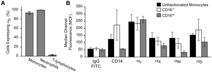
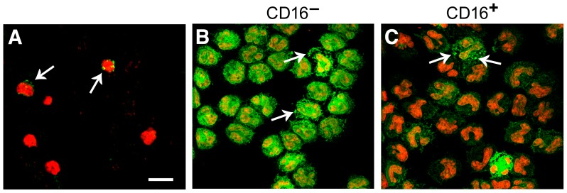
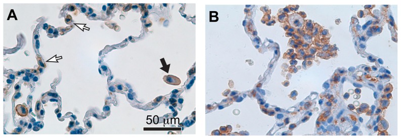
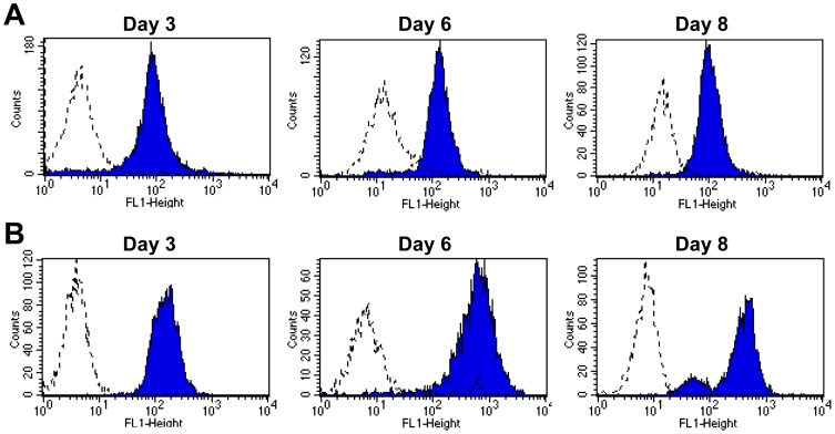
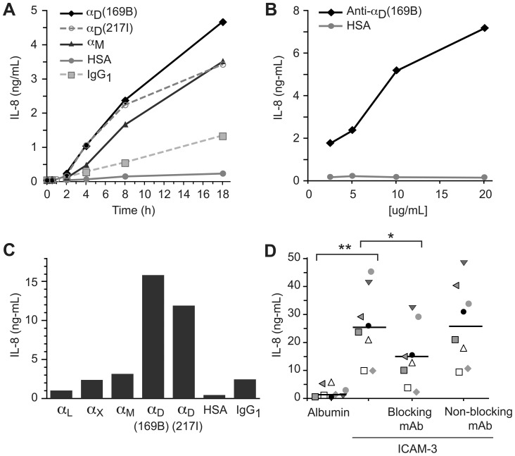
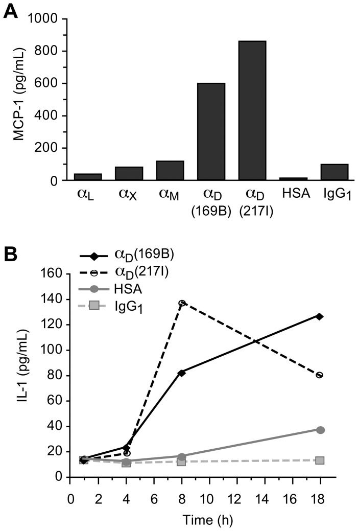
Integrin α(D)β(2) is the most recently identified member of the leukocyte, or β(2), subfamily of integrin heterodimers. Its distribution and functions on human leukocytes have not been clearly defined and are controversial. We examined these issues and found that α(D)β(2) is prominently expressed by leukocytes in whole blood from healthy human subjects, including most polymorphonuclear leukocytes and monocytes. We also found that α(D)β(2) is displayed by leukocytes in the alveoli of uninjured and inflamed human lungs and by human monocyte-derived macrophages and dendritic cells, indicating broad myeloid expression. Using freshly-isolated human monocytes, we found that α(D)β(2) delivers outside-in signals to pathways that regulate cell spreading and gene expression. Screening expression analysis followed by validation of candidate transcripts demonstrated that engagement of α(D)β(2) induces mRNAs encoding inflammatory chemokines and cytokines and secretion of their protein products. Thus, α(D)β(2) is a major member of the integrin repertoire of both circulating and tissue myeloid leukocytes in humans. Its broad expression and capacity for outside-in signaling indicate that it is likely to have important functions in clinical syndromes of infection, inflammation, and tissue injury.


Conflict of interest statement

Competing Interests: The authors have declared that no competing interests exist.

Figures

Figure 1
Figure 1. Integrin αDβ2 is highly expressed on myeloid leukocytes in human blood.
A. Whole venous blood from healthy volunteers was fixed and expression of αDβ2 on leukocyte subtypes was examined using mAb 169A (anti-αD) and FITC-conjugated mAbs against CD14, CD15, and CD3 as described in Materials and Methods. The percent of each cell type positive for αDβ2 is indicated by the bars. The error bars indicate the mean and SD of determinations using samples from four subjects. B. Monocytes were separated from venous blood of healthy volunteers and further separated into CD16+ and CD16 CD14+ subpopulations as described in Materials and Methods. Surface expression of αDβ2, αLβ2, αMβ2, and αXβ2 was examined by flow cytometry using FITC- or ALEXA-488-conjugated antibodies and isotype-matched IgG controls. Cells in each monocyte fraction were also stained for CD14. These data indicate means and standard deviations in results from 3 experiments using samples from different subjects.
Figure 2
Figure 2. Integrin αDβ2 is expressed by inflamed murine leukocytes and by unstimulated circulating human monocytes.
A. Leukocytes in whole cardiac blood from a mouse infected with the rodent malarial parasite Plasmodium berghei ANKA were stained for αDβ2 (arrows). B, C. Monocytes were first separated from an unfractionated mononuclear cell suspension from the peripheral blood of a healthy human volunteer and then further separated into CD16+ and CD16 subpopulations as described in Materials and Methods. The CD16 CD14+ (B) and CD16+ (C) monocyte preparations were then fixed, permeabilized, and stained for αD (green fluorescence) using anti-αD mAb 169A. Propidium iodide was used to identify nuclei. In additional experiments isotype-matched non-immune IgG was used as the first immunoglobulin in the staining procedure to control for mAb 169A (Figure S2). In both monocyte subsets, αDβ2 staining had a granular pattern and in some areas there were large clusters of the integrin that appeared to be on or near the surface (arrows). An additional experiment indicated that αDβ2 also clusters on human neutrophils (Figure S3).
Figure 3
Figure 3. Integrin αDβ2 is expressed by leukocytes in human lung.
A. Tissue samples from a human subject with normal lungs who underwent autopsy after fatal head injury were fixed and stained for αD with mAb 169A as outlined in Materials and Methods and . Microscopic evaluation revealed αDβ2 + macrophages in alveolar spaces (black arrow). There are also scattered αD + cells in alveolar walls (white arrows), which may be interstitial macrophages and/or dendritic cells. This image is representative of findings from analysis of lung tissue from three subjects who died without lung disease or injury. B. Autopsy samples from a patient who died with acute respiratory distress syndrome , were fixed, stained for αD, and examined by light microscopy. Numerous αDβ2 + macrophages were detected in alveolar spaces and walls. In some fields αDβ2 + neutrophils were also present (not shown). Lung samples from 3 patients who died with acute lung injury or ARDS as defined by consensus criteria were examined. We found αDβ2 + leukocytes in the alveoli in each case.
Figure 4
Figure 4. Human monocyte-derived macrophages and dendritic cells express integrin αDβ2 during differentiation in culture.
Unfractionated monocytes were separated from the venous blood of healthy subjects by positive selection and differentiated to (A) macrophages or (B) monocyte-derived dendritic cells using procedures and protocols outlined in Materials and Methods. MDM and MDDC were examined for expression of αDβ2 by flow cytometry on days 3, 6, and 8 in culture. These results are representative of findings in three experiments using cells from different subjects.
Figure 5
Figure 5. Engagement of integrin αDβ2 induces expression and release of IL-8 by human monocytes.
Unfractionated human monocytes were incubated in wells coated with immobilized mAb against αD, or in wells coated with HSA or IgG1 as control conditions, for various times. Supernatants were collected and analyzed for IL-8 by ELISA. In some experiments engagement of αDβ2 was compared to engagement of other leukocyte integrins using immobilized mAb against individual leukocyte integrin α subunits. A. Engagement of αDβ2 by immobilized anti-αD mAb induced time-dependent release of IL-8, whereas IL-8 secretion by monocytes incubated on HSA- or IgG1-coated control surfaces was much lower. In parallel, incubation of monocytes with LPS (100 µg/ml) in suspension induced release of IL-8 at 8 (1.1 ng/ml) and 18 hr (4.4 ng/ml) (not shown). Engagement of αMβ2 also induced time-dependent release of IL-8. B. Release of IL-8 triggered by engagement of αDβ2 on monocytes was dependent on the concentration of anti-αD mAb used to coat the wells. This figure indicates the results from an 8 hr incubation of monocytes on the triggering and control surfaces. C. Engagement of integrin αDβ2 was a potent stimulus for IL-8 secretion at 8 hr when immobilized mAb against αD were compared to immobilized mAb against other leukocyte integrin α subunits. Two activating anti-αD monoclonal antibodies, 169B and 217I, were examined in this experiment. The data in Panels A-C are individual determinations in single experiments. Data from additional experiments done at the 8 hr time point using monocytes from multiple different donors are shown in Tables S3 and S4. D. Wells were coated with human albumin or recombinant ICAM-3, monocytes were incubated in these wells for 18 hr. alone, in the presence of a blocking anti-αD mAb (mAb 240I), or in the presence of a non-blocking anti-αD monoclonal antibody (mAb 169A). IL-8 was then measured in the supernatants. The figure indicates the results of incubations with monocytes from 8 different subjects studied in 5 separate experiments on different days. Results from each subject are identified by a different symbol. The horizontal bars in the columns of data points indicate the means of determinations for each condition. The data were analyzed with Tukey's multiple comparison test and the Neuman-Kuels multiple comparison test, with similar results in each case. The significance values from the Neuman-Kuels analysis are shown (** = p<0.001; * = p<0.01). There was no difference in release of the IL-8 when monocytes were incubated with ICAM-3 alone versus incubation with ICAM-3 in the presence of the non-blocking mAb (p>0.05).
Figure 6
Figure 6. Engagement of integrin αDβ2 triggers expression of inflammatory chemokines and cytokines by human monocytes.
Integrin αDβ2 was engaged by immobilized activating mAb and supernatants for mediator analysis were collected as outlined in Figure 5. In (A) the concentration of anti-αD mAb, mAb against other leukocyte integrin α subunits, or control proteins used to coat the wells was 10 µg/ml, and in (B) the concentrations were 20 µg/ml. A. Engagement of integrin αDβ2 (8 hr) triggered release of MCP-1 that was much greater than that induced by immobilized mAb against other leukocyte integrin α subunits or release from monocytes incubated on control surfaces. Two activating anti-αD mAb, 169B and 217I, were studied. This result is representative of the pattern seen in eight separate experiments using monocytes from different donors, as shown in detail in Table S5. B. Engagement of integrin αDβ2 induced time-dependent release of IL-1β by monocytes. Two activating anti-αD mAb were examined, as in (A). In five additional experiments using monocytes from different donors, IL-1β protein was induced in monocyte lysates and released into the supernatants when integrin αDβ2 was engaged for 8 hr., and was greater than that in samples from monocytes incubated on control surfaces (Table S6).

Similar articles

Cited by

References

    1. Hynes RO (2002) Integrins: bidirectional, allosteric signaling machines. Cell 110:673–687. - PubMed
    1. Barczyk M, Carracedo S, Gullberg D (2010) Integrins. Cell Tissue Res 339:269–280. - PMC - PubMed
    1. Lowell CA, Mayadas TN (2012) Overview: studying integrins in vivo. Methods Mol Biol 757:369–397. - PMC - PubMed
    1. Harris ES, McIntyre TM, Prescott SM, Zimmerman GA (2000) The leukocyte integrins. J Biol Chem 275:23409–23412. - PubMed
    1. Evans R, Patzak I, Svensson L, De Filippo K, Jones K, et al. (2009) Integrins in immunity. J Cell Sci 122:215–225. - PubMed

Publication types