. 2015 Jan;125(1):105-16.
doi: 10.1172/JCI65654. Epub 2014 Nov 21.

Viral-mediated RdCVF and RdCVFL expression protects cone and rod photoreceptors in retinal degeneration

Viral-mediated RdCVF and RdCVFL expression protects cone and rod photoreceptors in retinal degeneration

Leah C Byrne et al. J Clin Invest. 2015 Jan.

Abstract

Alternative splicing of nucleoredoxin-like 1 (Nxnl1) results in 2 isoforms of the rod-derived cone viability factor. The truncated form (RdCVF) is a thioredoxin-like protein secreted by rods that promotes cone survival, while the full-length isoform (RdCVFL), which contains a thioredoxin fold, is involved in oxidative signaling and protection against hyperoxia. Here, we evaluated the effects of these different isoforms in 2 murine models of rod-cone dystrophy. We used adeno-associated virus (AAV) to express these isoforms in mice and found that both systemic and intravitreal injection of engineered AAV vectors resulted in RdCVF and RdCVFL expression in the eye. Systemic delivery of AAV92YF vectors in neonates resulted in earlier onset of RdCVF and RdCVFL expression compared with that observed with intraocular injection using the same vectors at P14. We also evaluated the efficacy of intravitreal injection using a recently developed photoreceptor-transducing AAV variant (7m8) at P14. Systemic administration of AAV92YF-RdCVF improved cone function and delayed cone loss, while AAV92YF-RdCVFL increased rhodopsin mRNA and reduced oxidative stress by-products. Intravitreal 7m8-RdCVF slowed the rate of cone cell death and increased the amplitude of the photopic electroretinogram. Together, these results indicate different functions for Nxnl1 isoforms in the retina and suggest that RdCVF gene therapy has potential for treating retinal degenerative disease.


Figures
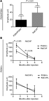
Figure 8
Figure 8. Injection of 7m8-scCAG-RdCVF rescues cones in P23H mice.
(A) Injections of 7m8-scCAG-RdCVF in homozygous P23H/P23H mice, a model of dominant RP, resulted in sustained amplitudes of the photopic ERG 1 month after injection (n = 6). (B) Time course of ERGs recorded from P23H/+ mice injected with 7m8-scCAG-RdCVF (n = 5) or 7m8-scCAG-RdCVFL (n = 6). In mice injected with RdCVF, ERG amplitudes were higher in treated eyes compared with those in control, PBS-injected, and contralateral eyes at 1 and 4 months, but not 6 months, after injection. In mice treated with 7m8-scCAG-RdCVFL, ERG amplitudes were not significantly different from those in sham-injected eyes at any of the time points measured.
Figure 7
Figure 7. Intraocular injection of 7m8-scCAG-RdCVF rescues cones.
(A) Photopic ERG amplitudes in rd10 animals injected at P15 with 7m8-GFP, PBS, 7m8-RdCVFL, 7m8-RdCVF, or a mix of 7m8-RdCVF plus 7m8-RdCVFL. Injection of 7m8-scCAG-RdCVFL resulted in a small but statistically insignificant increase in the amplitude of the photopic ERG b-wave compared with that measured in PBS- or GFP-injected eyes. Injection of 7m8-scCAG-RdCVF significantly increased the amplitude of the photopic ERG wave compared with that that of control eyes. Coinjection of 7m8-scCAG-RdCVF and 7m8-scCAG-RdCVFL resulted in greater rescue of the photopic ERG. n = 5 for all groups. Significance was determined using 1-way ANOVA with Tukey’s post-hoc multiple comparisons test. (B) Representative ERG traces from eyes injected with 7m8-GFP, 7m8-RdCVFL, 7m8-RdCVF, or a mixture of 7m8-RdCVF plus 7m8-RdCVFL. Arrows indicate the onset of stimulus. (C) Representative flicker ERGs recorded at 30 Hz of eyes injected with 7m8-RdCVF or a mix of 7m8-RdCVF plus 7m8-RdCVFL indicated improvement over ERGs of control 7m8-GFP–injected eyes. Marks on scale bar indicate 10-μV increments. (D) Flat-mounted retinae labeled with anti–S-opsin and –M/L-opsin antibodies revealed higher density of cones in RdCVF-treated eyes compared with those in GFP-expressing contralateral eyes. Lower images illustrate improved densities of surviving cones in the central retina near the optic nerve head (asterisk) in 7m8-RdCVF–injected eyes. (E) Quantification of cone densities revealed greater numbers of anti–S-opsin– and anti–M/L-opsin–labeled cones in treated eyes compared with those detected in contralateral GFP-expressing eyes (n = 4). *P < 0.05 and **P < 0.01 by 2-tailed, paired Student’s t test.
Figure 6
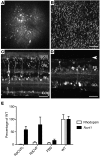
Figure 6. Expression profile of 7m8 after intravitreal injection at P15.
(A) Fundus image of P45 WT mouse illustrating strong GFP expression across the retina after intravitreal injection of 7m8-scCAG-GFP at P15. (B) Flat mount image of a retina expressing GFP, photoreceptor-side-up, showing GFP expression in outer segments of the photoreceptors. (C) Confocal stack of transverse sections showing expression of the reporter gene across all retinal layers in a WT mouse. (D) Confocal image of a P45 rd10 retina showing GFP expression in the GCL and INL and a loss of photoreceptors resulting in loss of the ONL (arrowhead). (E) qRT-PCR indicating near-WT levels of Nxnl1 mRNA expression from eyes injected with 7m8-scCAG-RdCVF and 7m8-scCAG-RdCVFL. GFP-injected eyes had markedly reduced Nxnl1 mRNA levels compared with levels in WT or AAV-injected eyes. Levels of rhodopsin mRNA were similarly low across conditions in rd10 eyes. mRNA was collected at P45 from 5 animals injected intravitreally at P15. Data represent the mean ± SD. n = 5 animals for each condition. Scale bars: 40 μm (B); 50 μm (C and D).
Figure 5
Figure 5. Effects of AAV92YF-mediated delivery of RdCVFL in dark-reared animals.
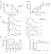
(A) Time course of the amplitude of the full-field scotopic ERG a-wave recorded in dark-reared rd10 mice injected with AAV92YF-RdCVFL (n = 5) or AAV92YF-scCAG-GFP (n = 5) revealed a delay in loss of retinal responses in treated animals. Four weeks after injection, a significant difference in the amplitude of the ERG a-wave was observed in RdCVFL-injected animals compared with that seen in GFP-injected mice. Five weeks after injection, both groups had progressively reduced a-wave amplitudes, and no difference in ERG amplitudes between the 2 groups was observed. Significance was determined using a repeated-measures 2-way ANOVA with Sidak’s multiple comparisons test. (B) A more detailed analysis of ERG traces recorded 4 weeks after injection of AAV92YF-RdCVFL or PBS revealed increased a-wave amplitudes over a range of flash intensities. Significance was determined using a 2-way ANOVA with post-hoc multiple comparisons test. (C) Representative ERG traces from RdCVFL-treated (left traces) or PBS-injected (right traces) animals illustrating increased amplitudes of the scotopic a-wave and b-wave 4 weeks after injection in treated mice. (D) Injection of AAV92YF-scCAG-RdCVFL resulted in an increase in the level of rhodopsin mRNA 4 weeks after injection compared with levels in untreated or GFP- or RdCVF-expressing eyes. Significance was determined by 1-way ANOVA with Tukey’s post-hoc multiple comparisons test. (E) TBARS assay revealed reduced levels of MDA, a marker for oxidative stress and lipid peroxidation, in retinae injected with AAV92YF-RdCVFL and collected 4 weeks after injection compared with MDA levels in AAV92YF-RdCVF, AAV92YF-GFP, or PBS-injected eyes (n = 3 for all groups). Data are presented as the percentage of change in MDA concentrations compared with those in untreated rd10 eyes ± SD. *P < 0.05; **P < 0.01.
Figure 4
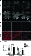
Figure 4. AAV92YF-mediated expression of RdCVF promotes cone survival.
(A) Flat mounts of retinae from animals injected with AAV92YF-scCAG-RdCVF and labeled with antibodies against M/L-opsin and S-opsin had greater numbers of cones than did PBS-injected mice. Top row: retinae from PBS-injected mice. Bottom row: retinae from AAV92YF-RdCVF–injected mice. (B) Higher-resolution images near the optic nerve (asterisk, upper right hand corner of each image) show increased density of M/L-opsin (red) or S-opsin (blue) in the central retina in RdCVF-injected animals. (C) Quantification of cones (n = 3) revealed an increase in the density of anti–L-opsin– and anti–S-opsin–labeled cones in retinae expressing RdCVF. Data represent the mean ± SD. **P < 0.01 by 2-tailed, unpaired Student’s t test.
Figure 3
Figure 3. Rescue of ERG amplitudes is dose dependent.
Representative traces (n = 3) are photopic ERGs from rd10 mice 1 month after injection with E+11 or E+12 particles. Arrows indicate the onset of the light flash.
Figure 2
Figure 2. Early delivery of RdCVF leads to sustained photopic ERG amplitudes.
(A) Photopic b-wave amplitudes were significantly higher in animals injected with AAV92YF-scCAG-RdCVF compared with those in mice injected with AAV92YF-scCAG-RdCVFL, AAV92YF-scCAG-GFP, or PBS. (B) Quantification (n = 5) of photopic ERG traces presented in A. **P < 0.01. Data are presented as the mean ± SD, and significance was determined using a 1-way ANOVA with Tukey’s post-hoc multiple comparisons test. (C) Representative flicker ERGs recorded from AAV92YF-scCAG-GFP–injected, AAV92YF-scCAG-RdCVF–injected, or WT mice (n = 5) illustrate improved amplitudes and more normal waveforms in treated animals. Marks on scale bar indicate 5-μV increments.
Figure 1
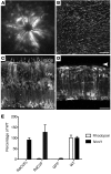
Figure 1. Expression profile of AAV92YF in the retina after systemic delivery at P1.
(A) Injection of AAV92YF-scCAG-GFP resulted in strong pan-retinal GFP expression, as shown in a fundus image of a P45 WT mouse. (B) Retinal flat mount showing large numbers of GFP-expressing photoreceptors in the central retina of a WT mouse 45 days after injection. (C) AAV92YF transduced cells in the GCL, INL, and ONL in a WT mouse. (D) Injection of AAV92YF led to gene expression across all layers of the retina in a P45 rd10 mouse. The ONL was significantly reduced at this time point (arrowhead). (E) qRT-PCR showing high levels of Nxnl1 mRNA from eyes injected with AAV92YF-scCAG-RdCVF or AAV92YF-scCAG-RdCVFL. PBS-injected animals had markedly reduced levels of Nxnl1 mRNA. Levels of rhodopsin expression were low across conditions in rd10 eyes. Eyes were collected at P35 from 3 animals injected in the tail vein at P1 and raised in a normal light/dark cycle. One eye was taken from each animal, and opposite eyes were used for the cone labeling and quantification shown in Figure 6. Scale bars: 40 μm (B); 50 μm (C and D).

References

    1. Buch H, et al. Prevalence and causes of visual impairment and blindness among 9980 Scandinavian adults: the Copenhagen City Eye Study. Ophthalmology. 2004;111(1):53–61. doi: 10.1016/j.ophtha.2003.05.010. - DOI - PubMed
    1. Daiger SP. RetNet: Summaries of Genes and Loci Causing Retinal Diseases. University of Texas Health Science Center Web site. https://sph.uth.edu/retnet/sum-dis.htm Updated October 14, 2014. Accessed October 28, 2014
    1. Bramall AN, Wright AF, Jacobson SG, McInnes RR. The genomic, biochemical, and cellular responses of the retina in inherited photoreceptor degenerations and prospects for the treatment of these disorders. Annu Rev Neurosci. 2010;33(1):441–472. doi: 10.1146/annurev-neuro-060909-153227. - DOI - PubMed
    1. Punzo C, Kornacker K, Cepko CL. Stimulation of the insulin/mTOR pathway delays cone death in a mouse model of retinitis pigmentosa. Nat Neurosci. 2009;12(1):44–52. doi: 10.1038/nn.2234. - DOI - PMC - PubMed
    1. Léveillard T, et al. Identification and characterization of rod-derived cone viability factor. Nat Genet. 2004;36(7):755–759. doi: 10.1038/ng1386. - DOI - PubMed

Publication types

MeSH terms