. 2015 Jun;45(6):1158-68.
doi: 10.1109/TCYB.2014.2346394. Epub 2014 Nov 14.

Label image constrained multiatlas selection

Label image constrained multiatlas selection

Pingkun Yan et al. IEEE Trans Cybern. 2015 Jun.

Abstract

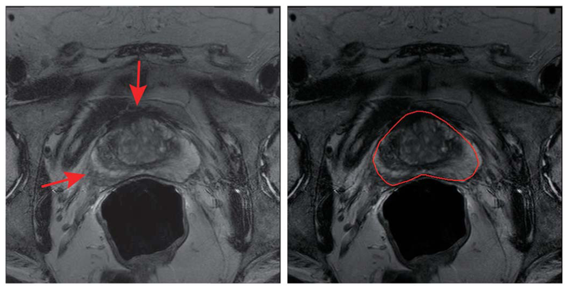
Multiatlas based method is commonly used in medical image segmentation. In multiatlas based image segmentation, atlas selection and combination are considered as two key factors affecting the performance. Recently, manifold learning based atlas selection methods have emerged as very promising methods. However, due to the complexity of prostate structures in raw images, it is difficult to get accurate atlas selection results by only measuring the distance between raw images on the manifolds. Although the distance between the regions to be segmented across images can be readily obtained by the label images, it is infeasible to directly compute the distance between the test image (gray) and the label images (binary). This paper tries to address this problem by proposing a label image constrained atlas selection method, which exploits the label images to constrain the manifold projection of raw images. Analyzing the data point distribution of the selected atlases in the manifold subspace, a novel weight computation method for atlas combination is proposed. Compared with other related existing methods, the experimental results on prostate segmentation from T2w MRI showed that the selected atlases are closer to the target structure and more accurate segmentation were obtained by using our proposed method.


Figures

Fig. 1.
Fig. 1.
Left: original MR image with two red arrows to illustrate the weakened boundary. Right: prostate segmentation delineated by an expert with red contour.
Fig. 2.
Fig. 2.
(a) Misleading manifold projection due to the influence of other anatomical structures in raw images. (b) Manifold projection constrained by the label images to reduce the influence and preserve the neighborhood structure.
Fig. 3.
Fig. 3.
Illustration of the mapping of combination weights. Assuming the raw images and the label image are embedded in the same manifold [34], the weight for combining label images combination can be computed by solving the problem of data points reconstruction in the lower-dimensional space.
Fig. 4.
Fig. 4.
Workflow of our proposed method. The upper row shows the process for analyzing the raw images of the atlases, and the lower row is the processing steps of the corresponding label images.
Fig. 5.
Fig. 5.
Randomly picked example with ground truth segmentation shown by the red surface. Along the atlas selection order from 1 to 10, the selected label images (green surfaces) are presented, respectively. The results, from top to bottom, are obtained by using NMI-based [11], LEAP [15], non-LICAS [16], LICAS-1, and LICAS-2, respectively.
Fig. 6.
Fig. 6.
Statistical performance of atlas selection on five methods along the atlas selection order from 1 to 10.
Fig. 7.
Fig. 7.
Some segmentation results of five methods. Yellow surfaces are the automatic segmentation results and the red surfaces are the ground truth.
Fig. 8.
Fig. 8.
(a) Average DSC of segmentation results of the five methods by varying the number of selected atlases from 1 to 10. (b) Comparison of the segmentation results by box-and-whisker diagrams.
Fig. 9.
Fig. 9.
Based on the same atlas selection results (LICAS-1), it compares the influence of different combination weights on the segmentation performance.
Fig. 10.
Fig. 10.
Spatial distribution of automatic segmentation errors on the Mollweide map based on the five methods.
Fig. 11.
Fig. 11.
Comparison of segmentation performance by varying the number of reduced dimension d. (a) LICAS-1. (b) LICAS-2.

References

    1. Yan P, Xu S, Turkbey B, and Kruecker J, “Discrete deformable model guided by partial active shape model for TRUS image segmentation,” IEEE Trans. Biomed. Eng, vol. 57, no. 5, pp. 1158–1166, May 2010. - PMC - PubMed
    1. Gao X, Wang B, Tao D, and Li X, “A relay level set method for automatic image segmentation,” IEEE Trans. Syst., Man, Cybern. B, Cybern, vol. 41, no. 2, pp. 518–525, April. 2011. - PubMed
    1. Pham D, Xu C, and Prince J, “Current methods in medical image segmentation,” Annu. Rev. Biomed. Eng, vol. 2, no. 1, pp. 315–337, 2000. - PubMed
    1. Guo Y, Zhan Y, Gao Y, Jiang J, and Shen D, “MR prostate segmentation via distributed discriminative dictionary (DDD) learning,” in Proc. IEEE 10th Int. Symp. Biomed. Imaging (ISBI), San Francisco, CA, USA, April. 2013. - PMC - PubMed
    1. Gao Y, Liao S, and Shen D, “Prostate segmentation by sparse representation based classification,” in Medical Image Computing and Computer-Assisted Intervention–MICCAI. Berlin, Germany: Springer, 2012, pp. 451–458. - PMC - PubMed

Publication types