. 2014 Nov 21;3(12):1252-61.
doi: 10.1242/bio.201410041.

Protein interference applications in cellular and developmental biology using DARPins that recognize GFP and mCherry

Affiliations

Protein interference applications in cellular and developmental biology using DARPins that recognize GFP and mCherry

Michael Brauchle et al. Biol Open. .

Abstract

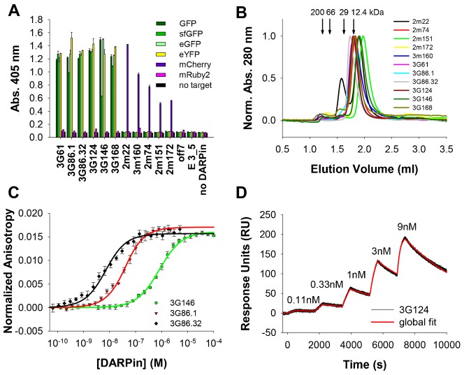
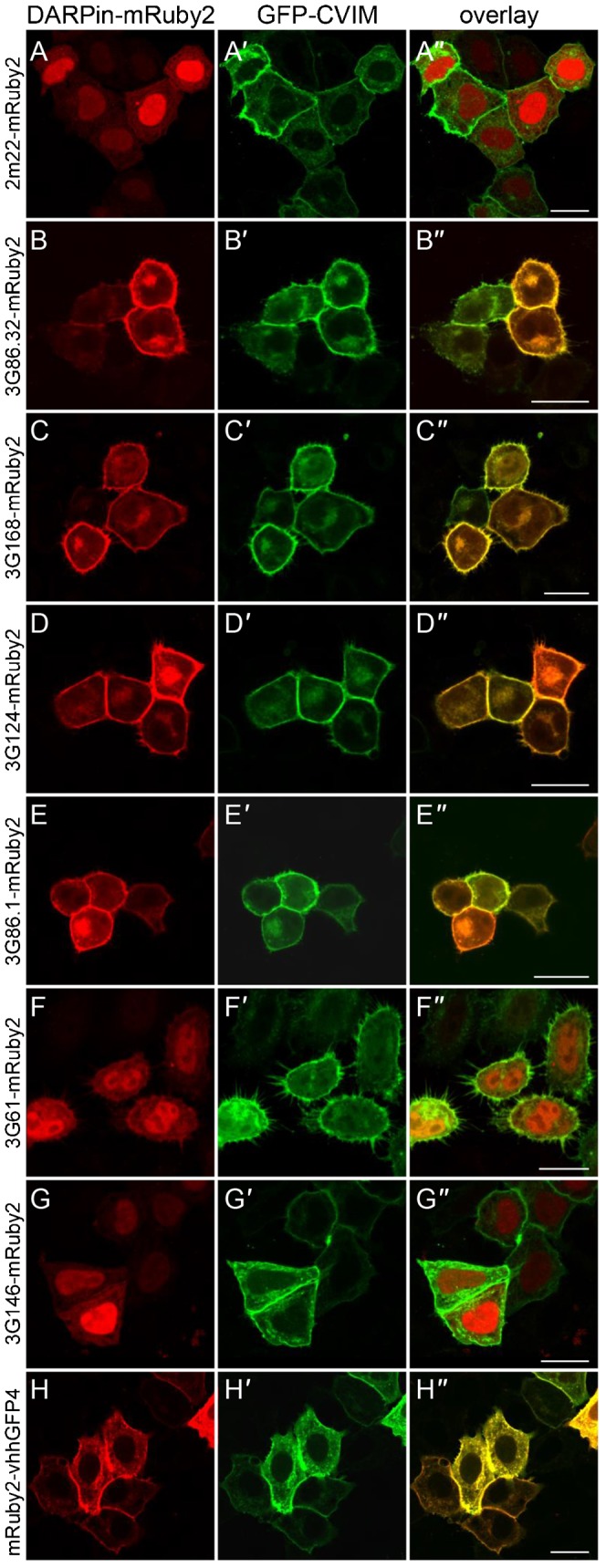
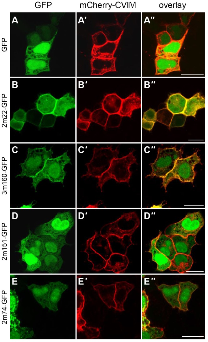
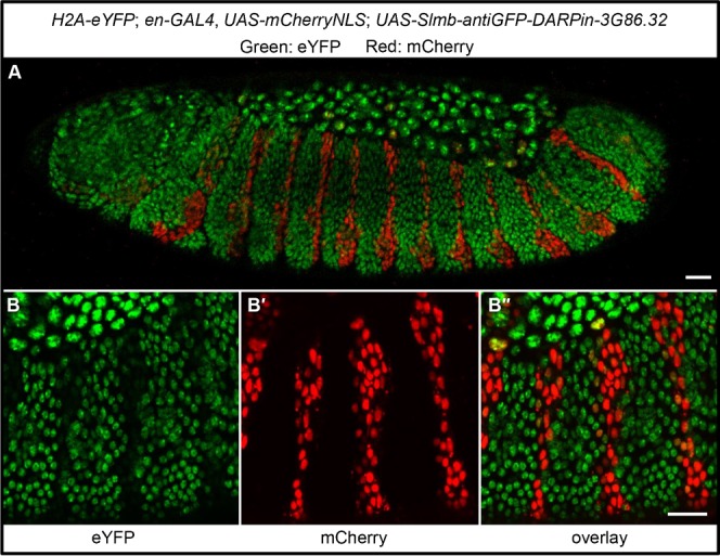
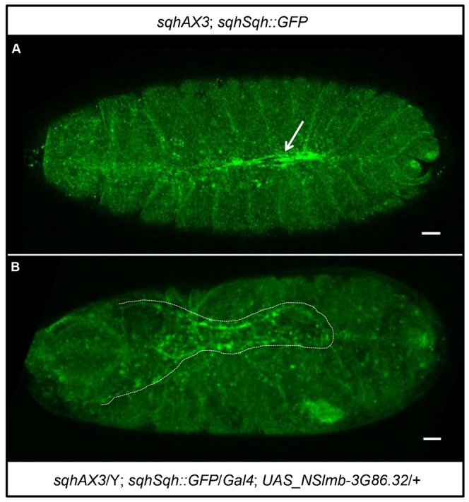
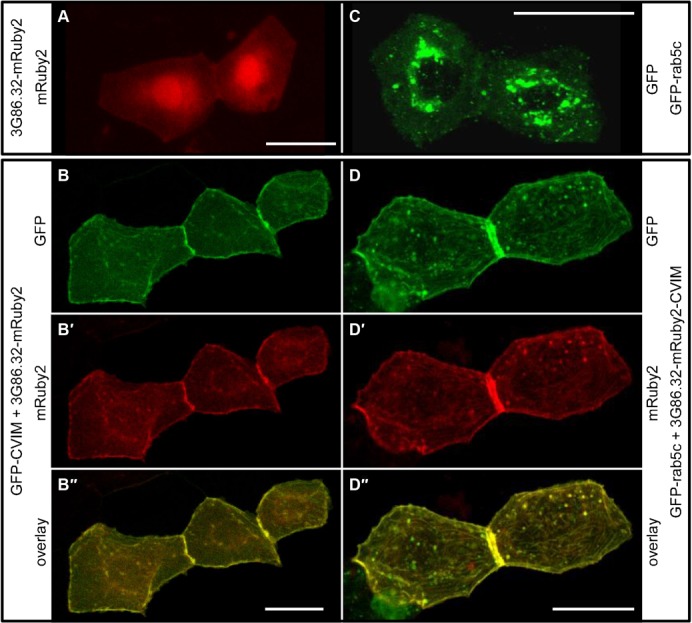
Protein-protein interactions are crucial for cellular homeostasis and play important roles in the dynamic execution of biological processes. While antibodies represent a well-established tool to study protein interactions of extracellular domains and secreted proteins, as well as in fixed and permeabilized cells, they usually cannot be functionally expressed in the cytoplasm of living cells. Non-immunoglobulin protein-binding scaffolds have been identified that also function intracellularly and are now being engineered for synthetic biology applications. Here we used the Designed Ankyrin Repeat Protein (DARPin) scaffold to generate binders to fluorescent proteins and used them to modify biological systems directly at the protein level. DARPins binding to GFP or mCherry were selected by ribosome display. For GFP, binders with KD as low as 160 pM were obtained, while for mCherry the best affinity was 6 nM. We then verified in cell culture their specific binding in a complex cellular environment and found an affinity cut-off in the mid-nanomolar region, above which binding is no longer detectable in the cell. Next, their binding properties were employed to change the localization of the respective fluorescent proteins within cells. Finally, we performed experiments in Drosophila melanogaster and Danio rerio and utilized these DARPins to either degrade or delocalize fluorescently tagged fusion proteins in developing organisms, and to phenocopy loss-of-function mutations. Specific protein binders can thus be selected in vitro and used to reprogram developmental systems in vivo directly at the protein level, thereby bypassing some limitations of approaches that function at the DNA or the RNA level.

Keywords: DARPin; GFP; Protein interference; mCherry.


Conflict of interest statement

Competing interests: The authors have no competing interests to declare. A.P. is a cofounder and shareholder of Molecular Partners AG, commercializing the DARPin technology.

Figures

Fig. 1.
Fig. 1.. Specificity, oligomeric state and affinity of anti-GFP and anti-mCherry DARPins.
(A) ELISA experiments show a high specificity of selected DARPins towards their cognate target and closely related proteins. Off7 is a control DARPin binding to maltose binding protein (MBP), E 3_5 is an unselected DARPin. All bars represent mean values of duplicates, error bars represent standard deviations. (B) Analysis of the oligomeric state by SEC shows that most DARPins are predominantly monomeric. Due to low extinction coefficients at 280 nm for the chromatograms of 2m22 and 2m74 the absorption at 230 nm is shown. Arrows indicate the elution volumes of the molecular weight standard with the respective MWs. (C) Example of FA assays of different DARPins binding to sfGFP. The solid line indicates a fit to a 1:1 binding model. Extracted KDs for all DARPins can be found in Table 1. (D) Example of a kinetic titration SPR experiment of 3G124 binding to GFP. The concentrations of the five DARPin injections are indicated in the graph. Fit to a global 1:1 kinetic titration binding model is indicated in red. Extracted association and dissociation rates and KDs for several DARPins are summarized in Table 1; additional sensograms are shown in supplementary material Fig. S4.
Fig. 2.
Fig. 2.. Binding of anti-GFP-DARPin-Ruby2 fusions to GFP in HeLa cells.
Shown are HeLa cells transiently overexpressing a DARPin-mRuby2 fusion protein (A–G) (H is an mRuby2-nanobody fusion) together with a GFP version tethered to the plasma membrane (GFP-CVIM, A′–H′). Overlap of fluorescent signal indicates binding of the respective anti-GFP-DARPin-mRuby2 fusion to GFP-CVIM, indicated by the yellow fluorescent signal at the plasma membrane. (A–A″) As expected, the anti-mCherry DARPin 2m22-mRuby2 fusion protein, which does not recognize GFP, is not recruited to the plasma membrane. (B–B″) 3G86.32-mRuby2, (C–C″) 3G168-mRuby2, (D–D″) 3G124-mRuby2 as well as (E–E″) 3G86.1-mRuby2 fusion proteins localize to the plasma membrane where they interact with GFP-CVIM. On the other hand, low affinity (F–F″) 3G61-mRuby2 and (G–G″) 3G146-mRuby2 fusion proteins localize to the cytoplasm and nucleus, indicating that they cannot interact sufficiently with GFP-CVIM anchored in the plasma membrane. (H–H″) Positive control mRuby2-VHH-GFP4 fusion protein localizes to the plasma membrane. Unprimed letters, mRuby2 channel; primed letters, GFP channel; double primed letters, overlay. Scale bars are 20 µm.
Fig. 3.
Fig. 3.. Binding of anti-mCherry-Darpin-GFP fusions to mCherry in HeLa cells.
Shown are HeLa cells transiently overexpressing GFP (A) or anti-mCherry-GFP fusion proteins (B–E) together with a mCherry version tethered to the plasma membrane (mCherry-CVIM, A′–E′). Overlap of fluorescent signal indicates binding of the respective anti-mCherry-DARPin-GFP fusion to mCherry-CVIM, indicated by the yellow fluorescent signal at the plasma membrane. (A–A″) As expected, the untethered GFP control does not change its subcellular localization upon co-expression with mCherry-CVIM and remains cytoplasmic and nuclear. (B–B″) 2m22-GFP re-localizes to the plasma membrane where it binds to mCherry-CVIM. (C–C″) The 3m160-GFP fusion protein results in some fluorescent signal at the plasma membrane indicating the weaker affinity to mCherry-CVIM. (D–D″) 2m151-GFP and (E–E″) 2m74-GFP fusion proteins are not significantly recruited to the plasma because of their only micromolar affinity. Unprimed letters, GFP channel; primed letters, mCherry channel; double primed letters, overlay. Scale bars are 20 µm.
Fig. 4.
Fig. 4.. Expression of a Slmb-anti-GFP-DARPin fusion in Drosophila embryos degrades eYFP.
(A) Shown is an embryo with the following genotype: H2A-eYFP; en-GAL4, UAS-mCherryNLS; UAS-Slmb-3G86.32. Nuclei that express the Slmb-anti-GFP-DARPin in the engrailed pattern are also expressing unfused mCherry. It can be seen that red cells lost the eYFP signal. (B–B″) Close-up of another embryo showing the channel-specific signal of eYFP (B), mCherry (B′) and the overlay (B″). Note again that cells expressing mCherry have strongly reduced H2A-eYFP signal, indicating efficient eYFP degradation due to the expression of a Slmb-anti-GFP-Darpin. Scale bars are 20 µm.
Fig. 5.
Fig. 5.. Tissue specific expression of a Slmb-anti-GFP-DARPin fusion in Drosophila phenocopies a non-muscle myosin II mutant phenotype.
(A) Shown is an embryo with the following genotype: sqhAX3; sqhSqh::GFP. The arrow points to the normal dorsal closure. (B) Shown is an embryo with the following genotype: sqhAX3/Y; sqhSqh::GFP/Gal4; NSlmb-3G86.32/+. The dotted line outlines the “dorsal open” phenotype exposing the amnioserosa. Scale bars are 20 µm.
Fig. 6.
Fig. 6.. anti-GFP-DARPin fusion proteins can relocalize fluorescent fusion proteins in D. rerio embryos.
(A,B) 3G86.2-mRuby2 binds GFP in living zebrafish embryos. (A) Control embryo showing the localization of 3G86.32-mRuby2 in two adjacent skin cells. (B–B″) In a zebrafish embryo co-expressing membrane-bound GFP-CVIM (B), 3G86.32-mRuby2 now localizes to the plasma membrane (B′) and shows a virtually complete co-localization with GFP-CVIM (B″). (C,D) membrane-anchored 3G86.32-mRuby2-CVIM recruits GFP-rab5c in living zebrafish embryos. (C) Control embryo showing the localization of GFP-rab5c in two adjacent skin cells. (D–D″) In a zebrafish embryo co-expressing membrane-bound 3G86.32-mRuby2-CVIM (D′), GFP-Rab5C also localizes to the plasma membrane (D) and shows a virtually complete co-localization with 3G86.32-mRuby2-CVIM (D″). Scale bars are 20 µm.

References

    1. Amstutz P., Binz H. K., Parizek P., Stumpp M. T., Kohl A., Grütter M. G., Forrer P., Plückthun A. (2005). Intracellular kinase inhibitors selected from combinatorial libraries of designed ankyrin repeat proteins. J. Biol. Chem. 280, 24715–24722 10.1074/jbc.M501746200 - DOI - PubMed
    1. Binz H. K., Plückthun A. (2005). Engineered proteins as specific binding reagents. Curr. Opin. Biotechnol. 16, 459–469 10.1016/j.copbio.2005.06.005 - DOI - PubMed
    1. Binz H. K., Stumpp M. T., Forrer P., Amstutz P., Plückthun A. (2003). Designing repeat proteins: well-expressed, soluble and stable proteins from combinatorial libraries of consensus ankyrin repeat proteins. J. Mol. Biol. 332, 489–503 10.1016/S0022-2836(03)00896-9 - DOI - PubMed
    1. Binz H. K., Amstutz P., Kohl A., Stumpp M. T., Briand C., Forrer P., Grütter M. G., Plückthun A. (2004). High-affinity binders selected from designed ankyrin repeat protein libraries. Nat. Biotechnol. 22, 575–582 10.1038/nbt962 - DOI - PubMed
    1. Bischof J., Maeda R. K., Hediger M., Karch F., Basler K. (2007). An optimized transgenesis system for Drosophila using germ-line-specific phiC31 integrases. Proc. Natl. Acad. Sci. USA 104, 3312–3317 10.1073/pnas.0611511104 - DOI - PMC - PubMed