. 2014 Nov 21;3(12):1217-23.
doi: 10.1242/bio.201410363.

Length-dependent anisotropic scaling of spindle shape

Affiliations

Length-dependent anisotropic scaling of spindle shape

Sarah Young et al. Biol Open. .

Abstract

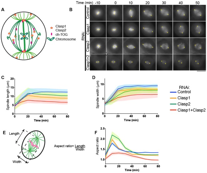
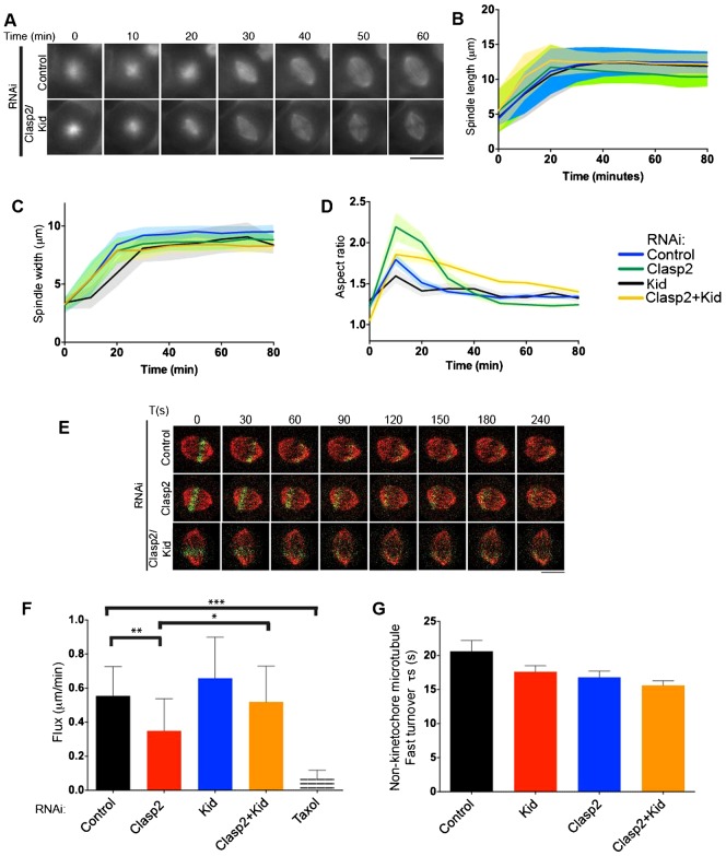
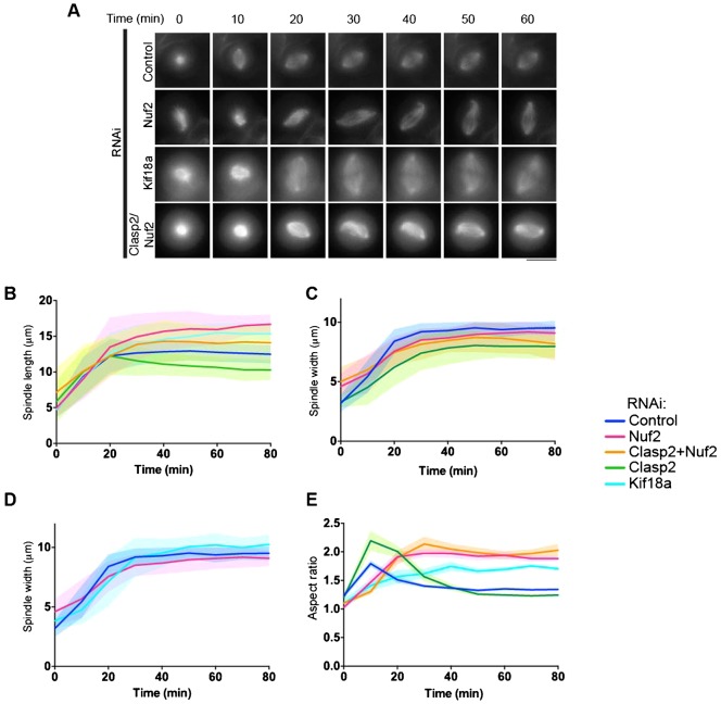
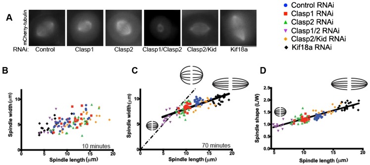
Spindle length varies dramatically across species and during early development to segregate chromosomes optimally. Both intrinsic factors, such as regulatory molecules, and extrinsic factors, such as cytoplasmic volume, determine spindle length scaling. However, the properties that govern spindle shape and whether these features can be modulated remain unknown. Here, we analyzed quantitatively how the molecular players which regulate microtubule dynamics control the kinetics of spindle formation and shape. We find that, in absence of Clasp1 and Clasp2, spindle assembly is biphasic due to unopposed inward pulling forces from the kinetochore-fibers and that kinetochore-fibers also alter spindle geometry. We demonstrate that spindle shape scaling is independent of the nature of the molecules that regulate dynamic microtubule properties, but is dependent on the steady-state metaphase spindle length. The shape of the spindle scales anisotropically with increasing length. Our results suggest that intrinsic mechanisms control the shape of the spindle to ensure the efficient capture and alignment of chromosomes independently of spindle length.

Keywords: Clasp; K-fiber; Microtubules; Mitosis; Spindle.


Conflict of interest statement

Competing interests: The authors have no competing interests to declare.

Figures

Fig. 1.
Fig. 1.. Clasp1 and Clasp2 depletion triggers a biphasic spindle assembly phase and changes in spindle geometry.
(A) Schematic diagram showing the localization of ch-TOG, Clasp1 and Clasp2 in a mitotic cell. The cell boundaries are represented in black, K-fibers, spindle microtubules and astral microtubules are represented in dark, medium and light green respectively. (B) Time-lapse imaging of U2OS cells expressing mCherry-tubulin after a STLC washout. Cells were treated with Control, Clasp1, Clasp2 and Clasp1/Clasp2 siRNA for 48 hours before imaging. The yellow asterisks represent the marking of the spindle pole used in the measurements of spindle length, using OMERO. (C,D,F) Graphs representing the average spindle length, width and aspect ratio and the corresponding SD, defined in panel E during elongation for each condition described in panel B. Scale bar: 10 µm.
Fig. 2.
Fig. 2.. The chromokinesin Kid opposes Clasp1 and Clasp2 during spindle formation.
(A) Time-lapse imaging of U2OS cells expressing mCherry-tubulin after a STLC washout. (B–D) Graphs representing the average spindle length, width and aspect ratio and the corresponding SD, during elongation for each condition described in panel B. (E) Time-lapse imaging of U2OS cells expressing mCherry-tubulin and PA-GFP-tubulin before and at 30 s intervals after photoactivation at 405 nm. (F,G) Quantification of microtubule poleward rate + SD and fast turnover + sem in cells treated with Control, Kid, Clasp2 or Clasp2/Kid siRNA. *, ** and *** represent a P<0.5, P<0.01 and P<0.001 respectively. Scale bars: 10 µm.
Fig. 3.
Fig. 3.. Clasp1 and Clasp2 act on kinetochore-fibers to stabilize the steady-state metaphase spindle.
(A) Time-lapse imaging of U2OS cells expressing mCherry-tubulin after a STLC washout, treated with indicated siRNA. (B–E) Graphs representing the average spindle length, width and aspect ratio and the corresponding SD during elongation for each condition described in panel A. Scale bar: 10 µm.
Fig. 4.
Fig. 4.. Spindle shape is modulated by regulators of microtubule dynamics.
(A) Representative images of spindles for cells depleted with indicated siRNA. Graphs representing spindle width versus spindle length for conditions in (B) 10 minutes and (C) 70 minutes after centrosome separation for conditions described above. The dashed line represents theoretical isotropic scaling. (D) Graph representing spindle aspect ratio versus spindle length for conditions in panel A. Raw data (118 points) were fitted to a linear regression function (R2 = 0.80). Scale bar: 10 µm.

References

    1. Al-Bassam J., Kim H., Flor-Parra I., Lal N., Velji H., Chang F. (2012). Fission yeast Alp14 is a dose-dependent plus end-tracking microtubule polymerase. Mol. Biol. Cell 23, 2878–2890 10.1091/mbc.E12-03-0205 - DOI - PMC - PubMed
    1. Allan C., Burel J. M., Moore J., Blackburn C., Linkert M., Loynton S., Macdonald D., Moore W. J., Neves C., Patterson A. et al. (2012). OMERO: flexible, model-driven data management for experimental biology. Nat. Methods 9, 245–253 10.1038/nmeth.1896 - DOI - PMC - PubMed
    1. Bertran M. T., Sdelci S., Regué L., Avruch J., Caelles C., Roig J. (2011). Nek9 is a Plk1-activated kinase that controls early centrosome separation through Nek6/7 and Eg5. EMBO J. 30, 2634–2647 10.1038/emboj.2011.179 - DOI - PMC - PubMed
    1. Bieling P., Kronja I., Surrey T. (2010). Microtubule motility on reconstituted meiotic chromatin. Curr. Biol. 20, 763–769 10.1016/j.cub.2010.02.067 - DOI - PubMed
    1. Blangy A., Lane H. A., d'Hérin P., Harper M., Kress M., Nigg E. A. (1995). Phosphorylation by p34cdc2 regulates spindle association of human Eg5, a kinesin-related motor essential for bipolar spindle formation in vivo. Cell 83, 1159–1169 10.1016/0092-8674(95)90142-6 - DOI - PubMed