. 2015 Jan 15;308(2):L147-57.
doi: 10.1152/ajplung.00108.2014.

Metabolic disturbances of the vitamin A pathway in human diaphragmatic hernia

Metabolic disturbances of the vitamin A pathway in human diaphragmatic hernia

Karen Coste et al. Am J Physiol Lung Cell Mol Physiol. .

Abstract

Congenital diaphragmatic hernia (CDH) is a common life-threatening congenital anomaly resulting in high rates of perinatal death and neonatal respiratory distress. Some of the nonisolated forms are related to single-gene mutations or genomic rearrangements, but the genetics of the isolated forms (60% of cases) still remains a challenging issue. Retinoid signaling (RA) is critical for both diaphragm and lung development, and it has been hypothesized that subtle disruptions of this pathway could contribute to isolated CDH etiology. Here we used time series of normal and CDH lungs in humans, in nitrofen-exposed rats, and in surgically induced hernia in rabbits to perform a systematic transcriptional analysis of the RA pathway key components. The results point to CRPBP2, CY26B1, and ALDH1A2 as deregulated RA signaling genes in human CDH. Furthermore, the expression profile comparisons suggest that ALDH1A2 overexpression is not a primary event, but rather a consequence of the CDH-induced lung injury. Taken together, these data show that RA signaling disruption is part of CDH pathogenesis, and also that dysregulation of this pathway should be considered organ specifically.


Figures

Fig. 1.
Fig. 1.
Overview of the retinoid signaling (RA) signaling pathway. Through a multistep cytosolic process, plasmatic retinol and β-carotene are converted into retinaldehyde and finally into retinoic acid. Retinol can be stored as retinyl ester. RA is transferred down to the nucleus where it binds to RA receptor dimers (RAR/RXR) and acts at short DNA sequences (RA response elements) to activate the transcription of target genes. Alternatively, intracellular RA can be catabolized through the cytochrome P-450 pathway. AKR1B1 and AKR1B10, aldo-keto reductase 1B1 and 1B10; ADH3 and ADH4, alcohol dehydrogenase 3 and 4; ALDH1A1, ALDH1A2, and ALDH1A3, aldehyde dehydrogenase 1 family, member A1 to A3 (also known as RALDH1-3); ALDH8A1, aldehyde dehydrogenase 8 family, member A1 (also known as RALDH4); b-ctn, β-carotene; BCMO1, β-carotene 15,15′-monooxygenase 1; BCO2, β-carotene oxygenase 2; CRABP1 and CRABP2, cellular RA binding protein 1 and 2; CRBP1 and CRBP2, cellular retinol binding protein 1 and 2; CYP26A1, CYP26B1, and CYP26C1, cytochrome P-450 A1, B1, and C1; DGAT, diacylglycerol acyl transferase; DHRS4 and DHRS9, short-chain dehydrogenase reductase 4 and 9; LRAT, lecithin retinol acyltransferase; RA, retinoic acid; Ral, retinaldehyde; RAR, retinoic acid receptor; RARE, retinoic acid response element; RBP4, retinol binding protein 4; RDH10, retinol dehydrogenase 10 (all-trans); RE, retinyl esters; REH, retinyl ester hydrolase; Rol, retinol; RXR, retinoid X receptor; STRA 6, stimulated by retinoic acid 6.
Fig. 2.
Fig. 2.
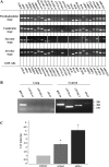
RA signaling components are expressed throughout the human lung development. A: representative qualitative PCR (qPCR) showing the expression timing of 25 RA signaling related genes at the pseudoglandular, canalicular, and saccular stages. Patterns in whole lungs are similar to the expression profile in the human alveolar epithelial cell line A549. Spaces demarcate independent gels. B: positive controls using tissue mix (liver, kidney, heart, and placenta) demonstrate that CY26B1, CYP26B1, and LRAT are not expressed in the human fetal lung. Samples were processed in parallel, and PCR products were controlled on different agarose gels. C: CAT gene reporter assay in A549 cells after 24 h of incubation with 1 μmol/l of retinol or RA. *P < 0.05; n = 4. RPLP0, ribosomal protein, large, P0.
Fig. 3.
Fig. 3.
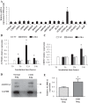
Diaphragmatic hernia disrupts RA signaling gene expression in human lung. A: relative mRNA expression of the RA signaling genes panel [congenital diaphragmatic hernia (CDH)/normal, respectively, n = 9 and n = 10] as measured by qPCR. CYP26B1 and CRBP2 are not expressed in CDH-injured lungs. B and C: bar chart showing, respectively, CYP26B1 and CRBP2 relative induction in A549 cells as measured by qPCR (n = 4) after treatment (6th, 24th, and 48th hours). White bar: no treatment (NT); gray bar: vehicle (DMSO); black bar (ATRA, 1 μmol/l). D: representative image of Western blotting with ALDH1A2 and GAPDH specific antibodies in one human CDH-injured lung and one normal human lung. According to the manufacturer's technical information, the ALDH1A2 antibody (sc-22591) detects two ALDH1A2 isoforms around 55 kDa. E: relative ALDH1A2 protein levels in normal human lungs (n = 4) and human CDH-injured lungs (n = 4). Results are corrected for GAPDH protein levels. *P < 0.05.
Fig. 4.
Fig. 4.
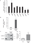
RA signaling genes are deregulated in animal CDH models. A: rat nitrofen CDH model; relative mRNA expression in lungs from nitrofen-exposed rats (n = 5) as measured by qPCR. B: rabbit surgical CDH model. Relative Aldh2A1 expression in control lungs (n = 4) and CDH-injured lungs (n = 4) as measured by qPCR. C: rabbit surgical CDH model; representative image of Western blotting with ALDH1A2 and GAPDH specific antibodies in one rabbit CDH-injured lung and one normal rabbit lung. D: rabbit surgical CDH model; relative ALDH1A2 lung protein levels in sham group (n = 4) and CDH group (n = 4). *P < 0.05; **P < 0.001.
Fig. 5.
Fig. 5.
Lung epithelial ALDH1A2 localization is not modified by rabbit surgically induced diaphragmatic hernia. Immunostained sections of human lung (AD: canalicular stage; I–L: saccular stage), rabbit lung (E–H: saccular stage; M–P: alveolar stage), and CDH-injured lung in rabbit (Q–T: saccular stage) showing the temporal and site-specific expression of ALDH1A2 in the epithelial cells from bronchi down to alveoli. Q–T: CDH-injured lungs in rabbits showing increased ALDH1A2 staining in epithelial cells, without modification of localization. Right: higher magnifications. Counterstaining: Hoechst; b, bronchi; s, saccule; a, alveoli. Scale bar: 100 μm.
Fig. 6.
Fig. 6.
Aldh1A2 is strongly expressed at the saccular stage, in both rabbit and human lungs. Relative Aldh1a2 mRNA expression in rabbit (A) and human lung (B), as measured by qPCR from the pseudoglandular to the alveolar or alternatively saccular stage (pseudoglandular stage: n = 4; the following stages: n = 3). *P < 0.05.

References

    1. Babiuk RP, Thebaud B, Greer JJ. Reductions in the incidence of nitrofen-induced diaphragmatic hernia by vitamin A and retinoic acid. Am J Physiol Lung Cell Mol Physiol 286: L970–L973, 2004. - PubMed
    1. Beurskens LWJE, Tibboel D, Lindemans J, Duvekot JJ, Cohen-Overbeek TE, Veenma DCM, de Klein A, Greer JJ, Steegers-Theunissen RPM. Retinol status of newborn infants is associated with congenital diaphragmatic hernia. Pediatrics 126: 712–720, 2010. - PubMed
    1. Brady PD, DeKoninck P, Fryns JP, Devriendt K, Deprest JA, Vermeesch JR. Identification of dosage-sensitive genes in fetuses referred with severe isolated congenital diaphragmatic hernia. Prenat Diagn 33: 1283–1292, 2013. - PubMed
    1. Brady PD, Srisupundit K, Devriendt K, Fryns JP, Deprest JA, Vermeesch JR. Recent developments in the genetic factors underlying congenital diaphragmatic hernia. Fetal Diagn Ther 29: 25–39, 2011. - PubMed
    1. Bustin SA, Benes V, Garson JA, Hellemans J, Huggett J, Kubista M, Mueller R, Nolan T, Pfaffl MW, Shipley GL, Vandesompele J, Wittwer CT. The MIQE guidelines: minimum information for publication of quantitative real-time PCR experiments. Clin Chem 55: 611–622, 2009. - PubMed

Publication types

MeSH terms

LinkOut - more resources