Review
. 2015 Feb 1;308(3):L229-52.
doi: 10.1152/ajplung.00238.2014. Epub 2014 Nov 21.

The role of inflammation in hypoxic pulmonary hypertension: from cellular mechanisms to clinical phenotypes

Affiliations
Review

The role of inflammation in hypoxic pulmonary hypertension: from cellular mechanisms to clinical phenotypes

Steven C Pugliese et al. Am J Physiol Lung Cell Mol Physiol. .

Abstract

Hypoxic pulmonary hypertension (PH) comprises a heterogeneous group of diseases sharing the common feature of chronic hypoxia-induced pulmonary vascular remodeling. The disease is usually characterized by mild to moderate pulmonary vascular remodeling that is largely thought to be reversible compared with the progressive irreversible disease seen in World Health Organization (WHO) group I disease. However, in these patients, the presence of PH significantly worsens morbidity and mortality. In addition, a small subset of patients with hypoxic PH develop "out-of-proportion" severe pulmonary hypertension characterized by pulmonary vascular remodeling that is irreversible and similar to that in WHO group I disease. In all cases of hypoxia-related vascular remodeling and PH, inflammation, particularly persistent inflammation, is thought to play a role. This review focuses on the effects of hypoxia on pulmonary vascular cells and the signaling pathways involved in the initiation and perpetuation of vascular inflammation, especially as they relate to vascular remodeling and transition to chronic irreversible PH. We hypothesize that the combination of hypoxia and local tissue factors/cytokines ("second hit") antagonizes tissue homeostatic cellular interactions between mesenchymal cells (fibroblasts and/or smooth muscle cells) and macrophages and arrests these cells in an epigenetically locked and permanently activated proremodeling and proinflammatory phenotype. This aberrant cellular cross-talk between mesenchymal cells and macrophages promotes transition to chronic nonresolving inflammation and vascular remodeling, perpetuating PH. A better understanding of these signaling pathways may lead to the development of specific therapeutic targets, as none are currently available for WHO group III disease.

Keywords: chronic nonresolving inflammation; fibroblasts; hypoxia; hypoxic pulmonary hypertension; inflammation; macrophages.


Figures

Fig. 1.
Fig. 1.
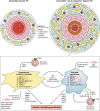
Inflammatory mechanisms involved in the initiation of hypoxia-induced pulmonary vascular remodeling. In hypoxic pulmonary hypertension (PH), the pulmonary artery (PA) undergoes changes in all three layers, including adventitial thickening with fibroblast proliferation and immune cell recruitment, medial thickening attributable mostly to smooth muscle cell hypertrophy, and intimal thickening attributable to subendothelial space thickening and endothelial cell hypertrophy and hyperplasia. These effects are mediated through the inflammatory cascade triggered by the combination of hypoxia and changes in flow/shear wall stress. These effects include generation of reactive oxygen species (ROS) through xanthine oxidoreductase (XOR), activation of NF-κB signaling, production of IL-6, and activation of hypoxia-inducible factor (HIF) and its downstream pathways. These pathways have important effects on each of the resident pulmonary vascular cells shown herein. PAEC, PA endothelial cell; SMC, smooth muscle cell; ECM, extracellular matrix.
Fig. 2.
Fig. 2.
Pulmonary vascular remodeling in brisket disease. Severe “out-of-proportion” brisket disease-induced pulmonary vascular remodeling is shown. The depiction is of a distal PA stained with Pentachrome from yearling steer that died of brisket disease despite relocation to a lower altitude. Strikingly, the neointima (neoint) is dramatically thickened and fibrotic, as highlighted by the blue color (ground substance), and causes luminal obliteration similar to lesions seen in irreversible PA hypertension. Also notice the dramatic neovascularization of all 3 layers of the vessel wall by vasa vasorum, as depicted by the arrowheads. The adventitial layer is dramatically increased in size and also fibrotic, as evidenced by collagen deposition (yellow). Adv, adventitia; M, media. Scale = 100 μm.
Fig. 3.
Fig. 3.
Cellular mechanisms involved in chronic nonresolving perivascular inflammation and irreversible pulmonary vascular remodeling. The transition from reversible pulmonary vascular remodeling to the irreversible remodeling seen in severe hypoxic PH involves increased thickening of all 3 layers of the vascular wall with fibrosis seen in both adventitial and intimal layers, leading to intimal occlusion. We hypothesize that these changes are mediated by hypoxia plus an as yet undefined “second hit,” which may include inflammation, infection, genetic, or environmental factors. Central to this hypothesis is the sentinel adventitial fibroblast, which undergoes important epigenetic changes that “lock” this cell into a proinflammatory and promitogenic phenotype. This epigenetically locked in fibroblast is characterized by increased HIF and NF-κB signaling and a shift to aerobic glycolysis, which triggers changes in micro-RNAs (mIRs), increased histone deacetylase (HDAC) activity, and increased DNA methylation. Collectively, these changes induce the fibroblast to recruit inflammatory monocytes, induce SMC hypertrophy and proliferation, and activate naïve monocytes into a unique proinflammatory and proremodeling phenotype via paracrine IL-6 and secreted metabolites, including lactate and succinate. This macrophage phenotype is characterized by signal transducer and activator of transcription 3 (STAT-3)-HIF-1-CCAAT/enhancer-binding protein-β (C/EBP-β) cosignaling, activation of NF-κB, and a shift to aerobic glycolysis. In a feed-forward loop, this macrophage can then further induce monocyte recruitment, macrophage activation, and SMC proliferation and activate naïve fibroblasts through various paracrine factors. Together, the fibroblast-macrophage signaling unit is able to perpetuate increased vascular remodeling and fibrosis through a persistent state of chronic nonresolving information. TIMPs, tissue inhibitors of metalloproteinases; MMPs, matrix metalloproteinases; SDF-1, stromal cell-derived factor 1; MCP-1, monocyte chemoattractant protein-1; CCR2, C-C chemokine receptor type 2; VWGF, vascular endothelial growth factor; ARG1, arginase 1.

References

    1. Aaronson PI, Robertson TP, Ward JP. Endothelium-derived mediators and hypoxic pulmonary vasoconstriction. Respir Physiol Neurobiol 132: 107–120, 2002. - PubMed
    1. Abe K, Toba M, Alzoubi A, Ito M, Fagan KA, Cool CD, Voelkel NF, McMurtry IF, Oka M. Formation of plexiform lesions in experimental severe pulmonary arterial hypertension. Circulation 121: 2747–2754, 2010. - PubMed
    1. Abud EM, Maylor J, Undem C, Punjabi A, Zaiman AL, Myers AC, Sylvester JT, Semenza GL, Shimoda LA. Digoxin inhibits development of hypoxic pulmonary hypertension in mice. Proc Natl Acad Sci USA 109: 1239–1244, 2012. - PMC - PubMed
    1. Adkins WK, Taylor AE. Role of xanthine oxidase and neutrophils in ischemia-reperfusion injury in rabbit lung. J Appl Physiol 69: 2012–2018, 1990. - PubMed
    1. Aggarwal S, Gross CM, Sharma S, Fineman JR, Black SM. Reactive oxygen species in pulmonary vascular remodeling. Compr Physiol 3: 1011–1034, 2013. - PMC - PubMed

Publication types

LinkOut - more resources