Review
. 2016 Feb;22(1):46-60.
doi: 10.1177/1073858414558065. Epub 2014 Nov 21.

Androgen Modulation of Hippocampal Structure and Function

Affiliations
Review

Androgen Modulation of Hippocampal Structure and Function

Sarah Atwi et al. Neuroscientist. 2016 Feb.

Abstract

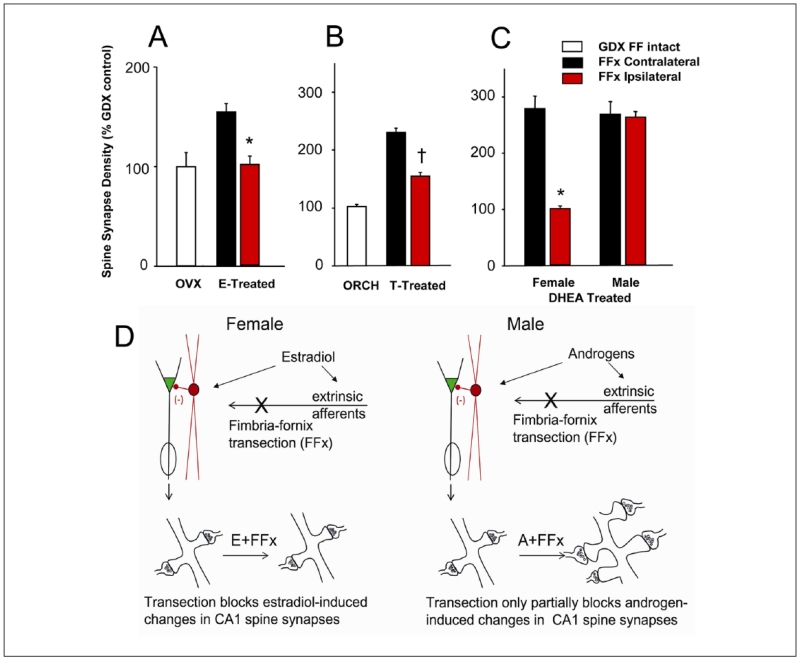
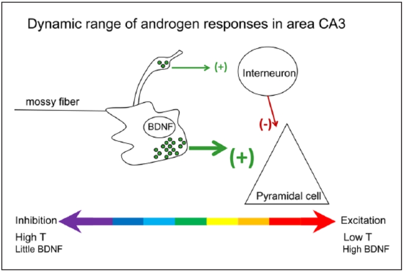
Androgens have profound effects on hippocampal structure and function, including induction of spines and spine synapses on the dendrites of CA1 pyramidal neurons, as well as alterations in long-term synaptic plasticity (LTP) and hippocampally dependent cognitive behaviors. How these effects occur remains largely unknown. Emerging evidence, however, suggests that one of the key elements in the response mechanism may be modulation of brain-derived neurotrophic factor (BDNF) in the mossy fiber (MF) system. In male rats, orchidectomy increases synaptic transmission and excitability in the MF pathway. Testosterone reverses these effects, suggesting that testosterone exerts tonic suppression on MF BDNF levels. These findings suggest that changes in hippocampal function resulting from declining androgen levels may reflect the outcome of responses mediated through normally balanced, but opposing, mechanisms: loss of androgen effects on the hippocampal circuitry may be compensated, at least in part, by an increase in BDNF-dependent MF plasticity.

Keywords: BDNF; CA3; hippocampus; mossy fibers; testosterone.


Figures

Figure 1
Figure 1
Effects on synapse formation in response to gonadal steroid administration of unilateral transection of the subcortical afferents to the hippocampus, by cutting the right fimbria/fornix (FF), are dependent on sex, as well as the nature of the gonadal steroid treatment. (A) Morphometric estimation of the density of spine synapses in the ipsilateral (red bars) and contralateral (black bars) CA1 stratum radiatum of unilaterally fimbria/fornix transected female rats. Rats were either ovariectomized (OVX) or OVX and treated with estradiol benzoate (2 × 10 μg, 24 hours apart, s.c.). FF transection had no effect on CA1 synapse density in OVX rats, but completely abolished the increase in synapse density induced by estradiol ipsilateral to the transection. *Significantly different from results on the contralateral side of the brain. (B) Density of pyramidal cell spine synapses in the CA1 stratum radiatum of intact male, orchidectomized (ORCH) and ORCH testosterone (T)-treated rats. Approximately equal spine synapse densities are present in the hippocampi of control males, ORCH T replaced males, and FF-transected T-replaced males contralateral to the FF transection. The spine synapse response to T is partially inhibited ipsilateral to FF transection. *Significantly different from control, intact males; Significantly different from both intact and ORCH males. (C) Spine synapses in the CA1 stratum radiatum of gonadectomized female and male rats after unilateral and dehydroepiandrosterone (DHEA) treatment. FF transection abolished the synaptic response to DHEA treatment ipsilateral to the transection in female, but not male, rats. In contrast to the results for T replacement, DHEA replacement in males was able to completely restore spine synapse density ipsilateral to FF transection. *Indicates a significant difference between the synapse densities in the ipsilateral and contralateral CA1. (D) Schematic illustration of the possible underlying mechanisms. In females, FF transection completely abolishes the ability of estradiol or the aromatizable androgen DHEA to increase CA1 spine synapse density, whereas in males the effects of androgen are only partially impaired by the elimination of subcortical afferents (adapted from data in Kovacs and others 2003; Leranth and others 2000; Mendell and others 2013).
Figure 2
Figure 2
Gonadectomized male rats exhibit increased immunoreactivity for BDNF protein in mossy fibers. Comparison of sham and gonadectomized male rats that were perfused and processed for immunocytochemistry using either a rabbit polyclonal BDNF antibody (a; antibody from Amgen Regeneron Partners), or a mouse monoclonal antibody to BDNF (b-d; antibody from Sigma). Arrows point to mossy fiber staining. DG = dentate gyrus; MOL = molecular layer; GCL = granule cell layer; HIL = hilus; SLM = stratum lacunosum moleculare; SR = stratum radiatum; SL = stratum lucidum; SP = stratum pyramidale; SO = stratum oriens. Calibration bars: a, b: 250 μm; c, d: 100 μm. Data from Skucas and others (2013).
Figure 3
Figure 3
Male gonadectomized rats exhibit increased mossy fiber transmission, paired pulse facilitation, and LTP. (A) Increasing stimulus strength to the mossy fibers produced field EPSPs (fEPSPs) with increasing slope and amplitude, recorded in stratum lucidum. Responses to stimuli are plotted for all sham (black circles) and gonadectomized (white) rats. Repeated-measures ANOVA showed that there was a significant effect of gonadectomy, and post hoc tests showed that most individual comparisons were significantly different (asterisks above the symbols). (B) Responses are shown for sham and gonadectomized rats with the recording electrode positioned in the pyramidal cell layer. Population spike amplitude was significantly larger in gonadectomized rats. (C) Recording electrodes were placed in stratum lucidum to examine paired pulse facilitation. Analysis of paired pulse facilitation with a range of interstimulus intervals from 20 to 200 ms showed that there was a significant effect of gonadectomy at intermediate intervals. (D) Comparison of LTP in gonadectomized (white circles) and sham rats (black circles) illustrates greater LTP in gonadectomized rats. Insets: representative examples of fEPSPs before and after LTP induction, with LTP evident only in the slice from the gonadectomized rat. Data from Skucas and others (2013).
Figure 4
Figure 4
Gonadectomized male rats exhibit mossy fiber sprouting. Examples of dynorphin immunocytochemistry in sham operated (A) and gonadectomized (B) male rats illustrate a novel band of immunoreactivity in stratum oriens (arrows) in gonadectomized rats. For abbreviations, see legend to Figure 2. Calibration bars: 500 μm (1); 100 μm (2), and 25 μm (3, 4). From Skucas and others (2013).
Figure 5
Figure 5
A schematic of the mossy fiber synapse shows a large bouton innervating a thorny excrescence of a CA3 pyramidal cell proximal apical dendrite. The large boutons have dense core vesicles that contain BDNF. A filamentous extension from the large bouton makes a smaller synaptic contact on a GABAergic neuron. Under normal conditions in the adult male rat, the mossy fiber system exerts a strong inhibitory tone because of the relatively low concentrations of BDNF (which normally facilitates glutamate release), the abundance of connections to the GABAergic interneurons, and high levels of the neurosteroid metabolite of testosterone, 5α-androstane-3α, 17β-diol, which facilitates GABA action at GABAA receptors. After gonadectomy, the mossy fiber pathway becomes more excitable, for two main reasons. First, there is a reduction in the effects of 5α-androstane-3α, 17β-diol at the GABAA receptor, because serum levels of its precursor (testosterone) fall. In addition, BDNF synthesis is increased while axonal sprouting occurs, so that more mossy fibers innervate area CA3 pyramidal cells (Skucas and others 2013).

References

    1. Acsady L, Kamondi A, Sik A, Freund T, Buzsaki G. GABAergic cells are the major postsynaptic targets of mossy fibers in the rat hippocampus. J Neurosci. 1998;18:3386–403. - PMC - PubMed
    1. Ahmadiani A, Mandgary A, Sayyah M. Anticonvulsant effect of flutamide on seizures induced by pentylenetetrazole: involvement of benzodiazepine receptors. Epilepsia. 2003;44:629–35. - PubMed
    1. Alibhai SM, Breunis H, Timilshina N, Marzouk S, Stewart D, Tannock I. Impact of androgen-deprivation therapy on cognitive function in men with nonmetastatic prostate cancer. J Clin Oncol. 2010;28:5030–7. others. - PubMed
    1. Alsemari A. Hypogonadism and neurological diseases. Neurol Sci. 2013;34:629–38. - PubMed
    1. Aubele T, Kaufman R, Montalmant F, Kritzer MF. Effects of gonadectomy and hormone replacement on a spontaneous novel object recognition task in adult male rats. Horm Behav. 2008;54:244–52. - PMC - PubMed

Publication types

LinkOut - more resources