. 2015 Jan 9;290(2):1256-68.
doi: 10.1074/jbc.M114.589838. Epub 2014 Nov 21.

MEF2 transcription factors regulate distinct gene programs in mammalian skeletal muscle differentiation

Affiliations

MEF2 transcription factors regulate distinct gene programs in mammalian skeletal muscle differentiation

Nelsa L Estrella et al. J Biol Chem. .

Abstract

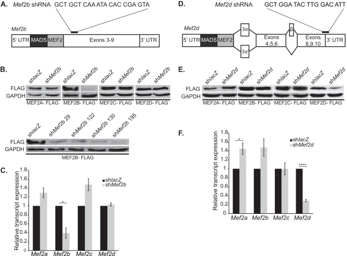
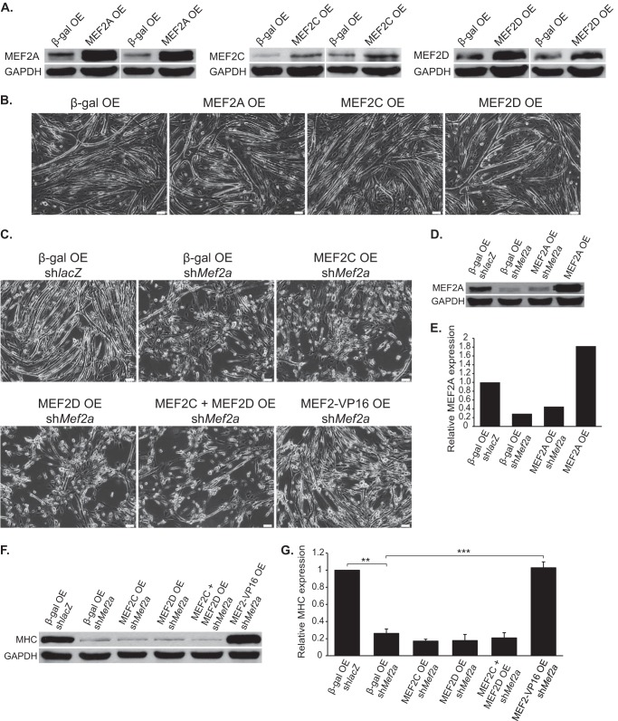
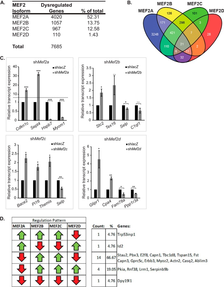
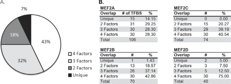
Skeletal muscle differentiation requires precisely coordinated transcriptional regulation of diverse gene programs that ultimately give rise to the specialized properties of this cell type. In Drosophila, this process is controlled, in part, by MEF2, the sole member of an evolutionarily conserved transcription factor family. By contrast, vertebrate MEF2 is encoded by four distinct genes, Mef2a, -b, -c, and -d, making it far more challenging to link this transcription factor to the regulation of specific muscle gene programs. Here, we have taken the first step in molecularly dissecting vertebrate MEF2 transcriptional function in skeletal muscle differentiation by depleting individual MEF2 proteins in myoblasts. Whereas MEF2A is absolutely required for proper myoblast differentiation, MEF2B, -C, and -D were found to be dispensable for this process. Furthermore, despite the extensive redundancy, we show that mammalian MEF2 proteins regulate a significant subset of nonoverlapping gene programs. These results suggest that individual MEF2 family members are able to recognize specific targets among the entire cohort of MEF2-regulated genes in the muscle genome. These findings provide opportunities to modulate the activity of MEF2 isoforms and their respective gene programs in skeletal muscle homeostasis and disease.

Keywords: Differentiation; Myogenesis; RNA Interference (RNAi); Transcription Factor; Transcriptomics.


Figures

FIGURE 1.
FIGURE 1.
Robust and specific knockdown of MEF2 proteins using shRNA adenoviruses. A and D, schematic representations of Mef2b (A) and Mef2d (D) transcripts. shRNA adenoviruses were generated to target the carboxyl-terminal region of the Mef2 transcripts (black bar) for knockdown in C2C12 myoblasts. The target sequences excluded regions containing alternatively spliced exons and were selected based on homology between mouse, human, and rat Mef2b or Mef2d sequences. B and E, Western blot analysis of shMef2b (B) and shMef2d (E) specificity against overexpressed MEF2 constructs in COS cells. Extracts for shMef2b knockdown of MEF2B-FLAG Western blot were cropped for image clarity (B, upper panel) but analyzed on the same gel (B, lower panel). shMef2b 195 (sequence shown in A) was used to generate adenovirus. C and F, quantitative RT analysis of endogenous Mef2b (C) and Mef2d (F) knockdown in C2C12 myotubes at differentiation day 3. The data are means ± S.E. *, p < 0.05; ****, p < 0.0001.
FIGURE 2.
FIGURE 2.
shRNA-mediated knockdown of MEF2 proteins in C2C12 myoblasts. C2C12 myoblasts were transduced with adenoviruses harboring shRNAs targeting Mef2a, -b, -c, or -d, or with an shRNA against lacZ as a negative control. A–C, knockdown of Mef2a (A), but not Mef2b, -c, or -d, resulted in impaired myotube formation and differentiation as shown by Western blot analysis of the muscle-specific marker myosin heavy chain (MHC) (B and C). D, combinatorial knockdown of Mef2a/b, Mef2a/c, and Mef2a/d failed to modulate the differentiation defect observed in the Mef2a knockdown alone. Additionally, simultaneous knockdown of Mef2b/c, Mef2b/d, Mef2c/d, and Mef2b/c/d failed to produce any overt morphological defects in C2C12 differentiation. E and F, Western blot analysis of myosin heavy chain (E) and accompanying densitometry (F) for the combinatorial knockdowns show that differentiation is only affected in MEF2A depleted cells. The data are means ± S.E. **, p < 0.01; ***, p < 0.001.
FIGURE 3.
FIGURE 3.
Overexpression and rescue of MEF2 in C2C12 myoblasts. A, Western blot analyses of C2C12 cells transduced with MEF2 isoform adenoviruses confirm an increase in MEF2 protein levels, relative to the β-gal control. OE, overexpression. B, overexpression of MEF2A, -C, or -D did not overtly modulate C2C12 myotube formation. Interestingly, overexpression of the MEF2D muscle-specific isoform did not result in enhanced myotube formation, as previously described. C, overexpression of MEF2-VP16, but not overexpression of MEF2C or -D alone or in combination, was able to rescue the differentiation defect observed in MEF2A depleted C2C12 myotubes. D, overexpression of MEF2A was unable to restore MEF2A to wild type levels, because the MEF2A cDNA encoded in this adenovirus is knocked down by shMef2a. E, quantification of MEF2A Western blot analysis. F, Western blot analysis of myosin heavy chain (MHC) expression demonstrates overexpression of MEF2-VP16, but not MEF2C or -D alone or in combination, was able to rescue impaired differentiation in MEF2A deficient cells. G, quantification of the myosin heavy chain expression Western blot analysis. The data are means ± S.E. **, p < 0.01; ***, p < 0.001.
FIGURE 4.
FIGURE 4.
Comparative analysis of MEF2 knockdown gene sets. Microarray analysis reveals that C2C12 cells are differentially sensitive to depletion of the four MEF2 isoforms. A, summary of total significantly dysregulated (q < 0.05) genes in each MEF2 isoform shRNA knockdown. B, a composite Venn diagram incorporating all overlapping gene sets as determined by the Tukey's honest significant difference post hoc test (q < 0.05). C, quantitative RT-PCR analysis of a subset of genes dysregulated in the Mef2 knockdown microarrays. Cdkn1c, cyclin-dependent kinase inhibitor 1c; Sept4, septin 4; Hspb7, heat shock protein family member 7; Myom1, myomesin 1; Stc2, stanniocalcin 2; Tex16, testis expressed gene 16; Selp, selectin, platelet; C1ql1, compliment component 1, q subcomponent-like 1; Bace2, beta-site app-cleaving enzyme 2; Pi16, peptidase inhibitor 16; Themis, thymocyte selection associated; Glipr1, GLI pathogenesis-related 1 (glioma); Cpa4, carboxypeptidase a4; Fam78a, family with sequence similarity 78; Ppp1r3a, protein phosphatase 1, regulatory (inhibitor) subunit 3a. The primer sequences are listed under “Experimental Procedures.” D, only five of the possible patterns of dysregulation are represented in the commonly dysregulated gene set. Of these patterns, the most prevalent group (66%) were genes that were down-regulated in MEF2A or MEF2D depletion and up-regulated in MEF2B or MEF2C depletion. Additionally, only a single gene, Dpy19l1 (DumPY19-like 1), was dysregulated in the same direction upon individual knockdown of each MEF2 factor. The data are means ± S.E. *, p < 0.05; **, p < 0.01; ***, p < 0.001; n.s., not significant.
FIGURE 5.
FIGURE 5.
Identification of distinct TF-binding sites associated with each MEF2 knockdown gene set. A, transcription factor-binding motif enrichment analysis was performed on the proximal promoter regions of each MEF2 gene set. Approximately 43% of binding motifs were shared by all four gene sets, 32% were shared by three MEF2 gene sets, 18% were shared by two MEF2 gene sets, and 7% are enriched only in genes regulated by a single MEF2 factor. B, breakdown of motif distribution by MEF2 isoform. MEF2A had the highest percentage of uniquely enriched motifs, and no unique motifs were identified for MEF2C-sensitive genes. TFBS, transcription factor binding site.

References

    1. Buckingham M., Rigby P. W. (2014) Gene regulatory networks and transcriptional mechanisms that control myogenesis. Dev. Cell 28, 225–238 - PubMed
    1. Braun T., Gautel M. (2011) Transcriptional mechanisms regulating skeletal muscle differentiation, growth and homeostasis. Nat. Rev. Mol. Cell Biol. 12, 349–361 - PubMed
    1. Black B. L., Olson E. N. (1998) Transcriptional control of muscle development by myocyte enhancer factor-2 (MEF2) proteins. Annu. Rev. Cell Dev. Biol. 14, 167–196 - PubMed
    1. Potthoff M. J., Olson E. N. (2007) MEF2: a central regulator of diverse developmental programs. Development. 134, 4131–4140 - PubMed
    1. Sandmann T., Jensen L. J., Jakobsen J. S., Karzynski M. M., Eichenlaub M. P., Bork P., Furlong E. E. (2006) A temporal map of transcription factor activity: Mef2 directly regulates target genes at all stages of muscle development. Dev. Cell 10, 797–807 - PubMed

Publication types

MeSH terms

Substances