. 2014 Nov 6;159(4):857-68.
doi: 10.1016/j.cell.2014.10.018.

Allosteric communication in the dynein motor domain

Affiliations

Allosteric communication in the dynein motor domain

Gira Bhabha et al. Cell. .

Abstract

Dyneins power microtubule motility using ring-shaped, AAA-containing motor domains. Here, we report X-ray and electron microscopy (EM) structures of yeast dynein bound to different ATP analogs, which collectively provide insight into the roles of dynein's two major ATPase sites, AAA1 and AAA3, in the conformational change mechanism. ATP binding to AAA1 triggers a cascade of conformational changes that propagate to all six AAA domains and cause a large movement of the "linker," dynein's mechanical element. In contrast to the role of AAA1 in driving motility, nucleotide transitions in AAA3 gate the transmission of conformational changes between AAA1 and the linker, suggesting that AAA3 acts as a regulatory switch. Further structural and mutational studies also uncover a role for the linker in regulating the catalytic cycle of AAA1. Together, these results reveal how dynein's two major ATP-binding sites initiate and modulate conformational changes in the motor domain during motility.


Figures
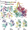
Figure 1
Figure 1. Crystal Structure of Yeast Dynein Motor Domain in the AMPPNP-Bound State
(A) Cartoon of the domain organization of the yeast dynein heavy chain and the crystal construct (a stalk-truncated motor domain harboring an E1849Q mutation at AAA1). (B) The overall structure of the motor domain-AMPPNP complex is shown in cartoon representation for the protein and in space-filling representation for AMPPNP ligands. (C) ATP-binding sites showing the density for AMPPNP molecules. The pink mesh shows an Fo – Fc omit map for AMPPNP, contoured at 3σ level. Side chains of the Walker A motif (K1802, T1803, T2425, K2080, T2081, and T2767), Walker B motif (D1848, Q1849, D2155, D2487, E2488, D2818, and E 2819), Sensor 1 (N1899, N2444, and T2890), Sensor 2 (R1971 and R2620), and R finger (R2209, R2552, R2911, and R3512) are shown in stick representation. (D and E) Binding to AMPPNP at AAA1 (D) and AAA3 (E) triggers closures of the AAA1–2 and AAA3–4 interfaces. The large domains are aligned. Arrows indicate a predominant rotation of the large and small domains of AAA1 and a rotation of the AAA3s-AAA4L interface. See text and Figure S1 for details. The color scheme is illustrated in (A).
Figure 2
Figure 2. Comparison of the Motor Domain Ring between Yeast apo, Yeast AMPPNP, and Dictyostelium ADP Crystal Structures
(A and B) Comparison of the two sides of the AAA ring in the indicated crystal structures. An upward movement of AAA2/3/4 toward the linker with the AMPPNP and ADP structures is observed (A), leading to a planar ring. In the ADP structure, AAA4 is lifted higher toward the linker. The line shows the common position of AAA1 in all structures. (B) An almost identical conformation of AAA5/6 for the apo and AMPPNP structures is observed, but the gap between AAA5 and AAA6 closes in the ADP structure (see box). Color coding of domains is the same as in Figure 1; structures are aligned on AAA1L. (C) Movements of the large domains of AAA4 and AAA5 relative to the linker (linker subdomains 1,2 aligned in these structures). The linker is docked to AAA5L, and AAA5/6 are in similar states in the apo and AMPPNP structures. However, in the ADP structure, the linker is undocked as a result of a movement of AAA5. See Figure S2 for supporting information. PDBs: 4AKG (Schmidt et al., 2012) for yeast apo and 3VKG (Kon et al., 2012) for Dictyostelium ADP. Note: subdomain 0 of the linker, the AAA5 extension, and C sequence were removed from the Dictyostelium structure for comparison with yeast.
Figure 3
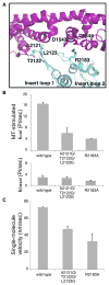
Figure 3. The Linker-Ring Interaction and Its Role in Dynein ATPase Activity and Motility
(A) The linker-AAA2 contacts in the yeast AMPPNP structure. (B) ATPase activity of dynein constructs in the absence (basal, bottom panel) or presence (MT-stimulated, top panel) of porcine MTs (see Experimental Procedures). The mean ± MT-stimulated kcat (mean ± SEM of four measurements from two independent protein preparations) is shown. (C) TMR-labeled, GST-dimerized yeast dynein constructs were tested for velocity in a single-molecule fluorescence motility assay (see Extended Experimental Procedures). The velocity (mean ± SEM of two independent protein preparations with n > 100 moving molecules each) is shown. See also Figure S3.
Figure 4
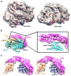
Figure 4. Cryo-EM Structure of Dynein in the Presence of ADP-AlF3 at an Average Resolution of ~10.5 Å
(A) Cryo-EM density (gray) with our AMPPNP crystal structure docked in. (B) Side view of the cryo-EM density and the docked AMPPNP crystal structure colored by domain. Density within 5 Å of each domain in the AMPPNP X-ray structure is colored. Insert, zoomed-in view of the contact between AAA2 loops and the linker; helices and loops fit reasonably well within the EM density. (C) Stereo view of density for linker docked to AAA5 and AAA1 is shown with the AMPPNP X-ray structure docked in. Representative data, the reconstruction colored by local resolution, other 3D classes, and negative-stain reconstructions for a construct containing the full stalk and MTBD are shown in Figure S4.
Figure 5
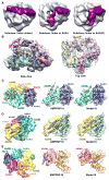
Figure 5. Cryo-EM Structure of Dynein in the Presence of ADP-Vanadate at an Average Resolution of ~9 Å
(A) 3D classes for cryo-EM data of dynein in the presence of ADP-vanadate: unbent linker (~39% particles), linker to AAA4 (~36% particles), and linker to AAA3/2 (~25% particles). The last subclass could be refined to the highest resolution, as shown in (B)–(E). (B) ADP-vanadate cryo-EM density fit with our model, which was generated from simultaneously fitting each s and L AAA subdomain into the density as rigid bodies in UCSF Chimera. (C–E) The large domains of the AAA ring, colored by domain, are shown on the left to provide a reference orientation for the fits of the cryo-EM electron density with the AMPPNP X-ray structure (middle) or the ADP-vanadate model (right). Domain motions of AAA2-AAA1 (C), AAA4-AAA3 (D), and AAA6-AAA5 (E) between the AMPPNP and ADP-vanadate states are shown. Representative data, the reconstruction colored by local resolution, 2D class averages, supporting 3D reconstructions from negative-stain EM data, and stereo views of Apo, AMPPNP, and the model fit in cryo-EM density as well as negative-stain data for similar complexes are shown in Figure S5.
Figure 6
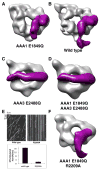
Figure 6. Blocking ATP Hydrolysis in AAA3 Prevents the Linker Conformational Change
(A–D) Negative-stain reconstructions for (A) AAA1 E1849Q, (B) wild-type, (C) AAA3 E2488Q, and (D) AAA1/AAA3 double Walker B mutant (E1849Q/E2488Q) dyneins. The dyneins were incubated with MgATP (5 mM) prior to negative staining. The electron density for the linker was clearly visible and is colored magenta. (E) Representative kymographs for single-molecule motility assays showing no detectable motility of the AAA2 R finger mutant (R2209A) (top panel) and microtubule-stimulated ATPase activity (bottom panel) for wild-type and the R2209A mutation (mean ± SEM of two independent protein preparations). (F) Negative-stain reconstruction for E1849Q/R2209A dynein in the presence of 5 mM ATP. Representative micrographs, additional 3D classes, and comparison with ADP instead of ATP for wild-type and the AAA1 E1849Q mutant are shown in Figure S6.
Figure 7
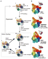
Figure 7. A Model for Structural Changes during Dynein’s ATPase Cycle
(A) The actively cycling states of the dynein motor are boxed (III, IV, and V), and repressed states are shown outside the box (I and II). Beginning with state III, ATP (“T”) binding to AAA1 results in the closures AAA1–2, which triggers a series of domain movements around the ring and closure of the AAA5–6 interface; movement of AAA5 results in linker detachment from AAA5 and a bent conformation of the linker. After phosphate release from AAA1, the linker straightens (the proposed power stroke) but remains undocked (IV). Linker docking to AAA5 promotes the further opening of AAA1–2 and ADP (“D”) release from AAA1, returning it to an apo state at AAA1 to begin a new cycle (V). If ADP is released (broken line from V) and ATP rebinds at AAA3 (II), the motor returns to the repressed state. See Discussion for more details. We denote the nucleotide state of AAA4 as “T/D” because our present model dos not incorporate a nucleotide-specific role at this site. A subtle modulatory role is possible, as a mutation blocking nucleotide hydrolysis at AAA4 produces a modest decrease in velocity and increase in processivity (Cho et al., 2008). (B) Surface representation of the AAA ring in yeast apo (PDB code 4AKG; Schmidt et al., 2012), yeast AMPPNP/ADP-AlF3 (our data), yeast ADP-vanadate (our data), and Dictyostelium ADP (PDB code 3VKG; Kon et al., 2012) used to synthesize the model presented in (A). We illustrate a model based on the Dictyostelium ADP X-ray structure, as a crystal structure for yeast ADP has not been obtained. Although the yeast ADP structure may differ it some details from Dictyostelium, the yeast ADP EM structure also clearly exhibits a “post-power-stroke” extended linker conformation (Figure S6F). Insets highlight the linker position in each state based on our EM data. See also Figure S7. The structural transitions in the dynein cycle can be viewed in Movie S1.

References

    1. Adams PD, Grosse-Kunstleve RW, Hung LW, Ioerger TR, McCoy AJ, Moriarty NW, Read RJ, Sacchettini JC, Sauter NK, Terwilliger TC. PHENIX: building new software for automated crystallographic structure determination. Acta Crystallogr D Biol Crystallogr. 2002;58:1948–1954. - PubMed
    1. Babst M, Wendland B, Estepa EJ, Emr SD. The Vps4p AAA ATPase regulates membrane association of a Vps protein complex required for normal endosome function. EMBO J. 1998;17:2982–2993. - PMC - PubMed
    1. Baker TA, Sauer RT. ClpXP, an ATP-powered unfolding and protein-degradation machine. Biochim Biophys Acta. 2012;1823:15–28. - PMC - PubMed
    1. Burgess SA, Knight PJ. Is the dynein motor a winch? Curr Opin Struct Biol. 2004;14:138–146. - PubMed
    1. Burgess SA, Walker ML, Sakakibara H, Knight PJ, Oiwa K. Dynein structure and power stroke. Nature. 2003;421:715–718. - PubMed

Publication types

MeSH terms