. 2014 Nov 6;159(4):884-95.
doi: 10.1016/j.cell.2014.09.055. Epub 2014 Oct 30.

Embryonic development following somatic cell nuclear transfer impeded by persisting histone methylation

Affiliations

Embryonic development following somatic cell nuclear transfer impeded by persisting histone methylation

Shogo Matoba et al. Cell. .

Abstract

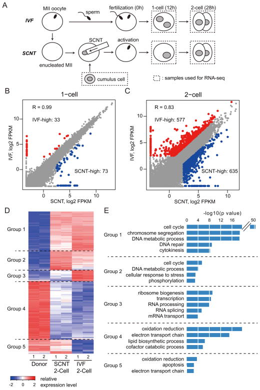
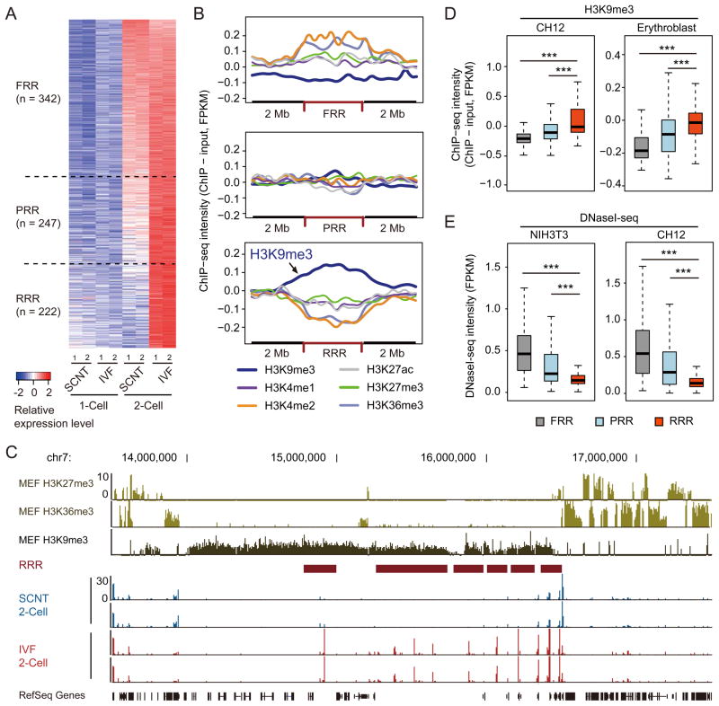
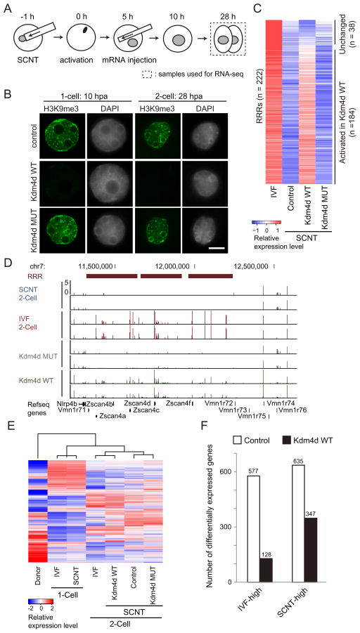
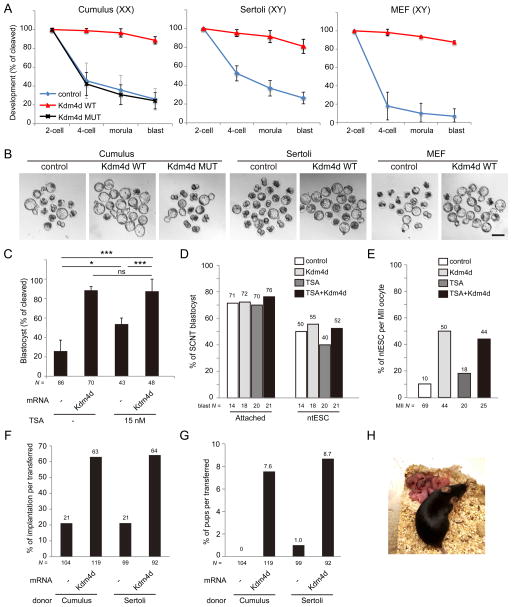
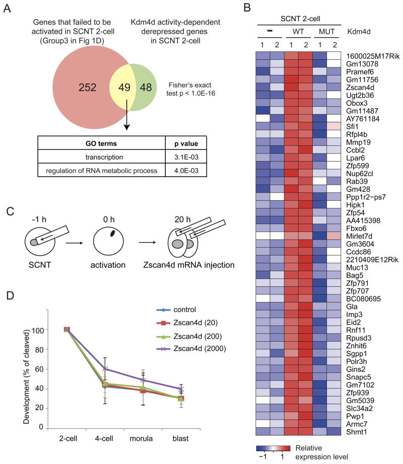
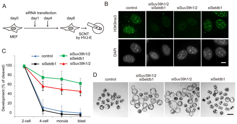
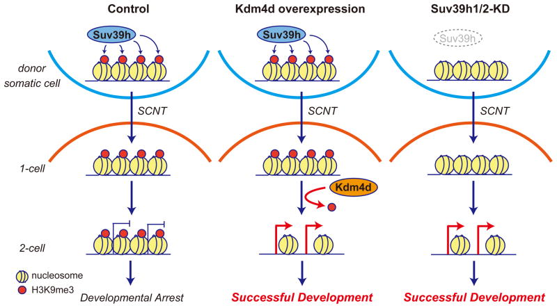
Mammalian oocytes can reprogram somatic cells into a totipotent state enabling animal cloning through somatic cell nuclear transfer (SCNT). However, the majority of SCNT embryos fail to develop to term due to undefined reprogramming defects. Here, we identify histone H3 lysine 9 trimethylation (H3K9me3) of donor cell genome as a major barrier for efficient reprogramming by SCNT. Comparative transcriptome analysis identified reprogramming resistant regions (RRRs) that are expressed normally at 2-cell mouse embryos generated by in vitro fertilization (IVF) but not SCNT. RRRs are enriched for H3K9me3 in donor somatic cells and its removal by ectopically expressed H3K9me3 demethylase Kdm4d not only reactivates the majority of RRRs, but also greatly improves SCNT efficiency. Furthermore, use of donor somatic nuclei depleted of H3K9 methyltransferases markedly improves SCNT efficiency. Our study thus identifies H3K9me3 as a critical epigenetic barrier in SCNT-mediated reprogramming and provides a promising approach for improving mammalian cloning efficiency.


Figures

Figure 1
Figure 1. Abnormal gene expression of SCNT embryos at the 1- and 2-cell stage
(A) Schematic illustration of the experimental approach. Samples used for RNA-seq are marked by dashed rectangles. (B, C) Scatter plots comparing gene expression levels between IVF and SCNT embryos at the 1-cell stage (B) and the 2-cell stage (C). Genes expressed higher in IVF embryos (FC > 3.0, IVF-high) and higher in SCNT embryos (FC > 3.0, SCNT-high) are colored with red and blue, respectively. (D) Heatmap illustration showing differentially expressed genes (DEGs) (FC > 5.0, FPKM > 5 in each replicates) obtained by a pairwise comparison between donor cumulus cells, IVF 2-cell and SCNT 2-cell embryos. A total of 3775 DEGs are classified into 5 groups by unsupervised hierarchical clustering. (E) Gene ontology analysis of the 5 groups classified in (D). See also Figure S1.
Figure 2
Figure 2. Reprogramming resistant regions (RRRs) are enriched for H3K9me3 in somatic cells
(A) Heatmap illustration of the transcripts of IVF and SCNT embryos. Each tile represents an average of peaks within the region obtained by sliding-window analysis. Shown are the 811 regions that are activated from 1-cell (12 h) to 2-cell (28 h) stage IVF embryos compared to cumulus derived SCNT embryos. These regions were classified into three groups based on the fold-change (FC) in transcription levels between SCNT- and IVF 2-cell embryos. FRRs, PRRs, and RRRs indicate fully reprogrammed regions (FC <= 2), partially reprogrammed regions (2 < FC <= 5) and reprogramming resistant regions (FC > 5), respectively. (B) The average ChIP-seq intensity of six histone modifications in MEF cells are shown within FRR, PRR and RRR compared with 2 MB flanking regions. Reads counts are normalized by input, total mapped reads and region length. (C) Representative genome browser view of RRRs on chromosome 7. (D, E) Box plots comparing the average intensity of H3K9me3-ChIP-seq (D) or DNaseI-seq (E) within FRR, PRR and RRR in different somatic cell types. ChIP-seq and DNaseI-seq datasets shown in (B–E) were obtained from ENCODE projects (Bernstein et al., 2012; The Encode Consortium Project, 2011). See also Figure S2.
Figure 3
Figure 3. Injection of Kdm4d mRNA removes H3K9me3 of transferred somatic cells and derepresses silenced genes in 2-cell SCNT embryos
(A) Schematic illustration of the experimental procedure. SCNT embryos derived from cumulus cells were injected with wild-type or a catalytic defective Kdm4d mRNA at 5 hours post activation (hpa). Samples used for RNA-seq are marked by dashed rectangles. (B) Representative nuclear images of 1-cell and 2-cell stage SCNT embryos stained with anti-H3K9me3 and DAPI. Shown in each panel is a nucleus of a single blastomere. Scale bar, 10 μm. (C) Heatmap comparing transcription levels of the 222 RRRs at the 2-cell stage. The expression level of 184 out of the 222 RRRs are significantly (FC > 2) increased in response to wild-type, but not the catalytic mutant, Kdm4d injection. (D) A genome browser view of an example of RRRs on chromosome 7. (E) Hierarchical clustering of all samples used in this study. Note that 2-cell SCNT embryos injected with wild-type Kdm4d were clustered together with 2-cell IVF embryos based on their transcriptome analysis. (F) Bar graph illustrates reduced number of differentially expressed genes (FC > 3) between IVF and SCNT 2-cell embryos after Kdm4d injection. See also Figure S3.
Figure 4
Figure 4. Injection of Kdm4d mRNA improves developmental potential of SCNT embryos
(A) Kdm4d mRNA injection greatly improves preimplantation development of SCNT embryos derived from cumulus cells, Sertoli cells and MEF cells. Shown is the percentage of embryos that reaches the indicated stages. XX and XY indicate the sex of donor mice. Error bars indicate s.d. (B) Representative images of SCNT embryos after 120 hours of culturing in vitro. Scale bar, 100 μm. (C) Kdm4d mRNA injection has additional effect over the treatment with Trichostatin A (TSA; 15 nM). Shown is the percentage of embryos that reached the blastocyst stage at 96 hpa. * P < 0.05, ** P < 0.01, *** P < 0.001. ns, not significant. (D) Bar graph showing the efficiency of attachment to the feeder cells and ntESC derivation of SCNT blastocysts. (E) Bar graph showing the efficiency of ntESC derivation. The efficiency was calculated based on the total number of MII oocytes used for the generation of SCNT embryos. (F, G) Implantation rate (F) and birth rate (G) of SCNT embryos examined by caesarian section on E19.5. (H) An image of an adult female mouse derived by SCNT of a cumulus cell with Kdm4d mRNA injection and its pups generated through natural mating with a wild-type male. See also Tables S1–3.
Figure 5
Figure 5. Candidate genes responsible for the poor developmental phenotype of SCNT embryos
(A) Venn diagram showing the overlap between the genes that failed to be activated in SCNT 2-cell embryos (Group3 in Figure 1D) and Kdm4d enzyme activity-dependently derepressed genes in SCNT 2-cell embryos. GO enrichment analysis was performed on the 49 overlap genes. (B) Heatmap showing the expression pattern of 49 overlap genes in (A). (C) Schematic illustration of the experimental procedure. Zscan4d mRNA was injected into both of 2-cell blastomeres of SCNT embryos at 20 hpa (early 2-cell stage). (D) Preimplantation development rate of SCNT embryos injected with Zscan4d mRNA at 0, 20, 200 or 2000 ng/μl. Error bars indicate s.d. of three biological replicates. See also Figure S4 and Table S4.
Figure 6
Figure 6. Suv39h1/2 is responsible for the establishment of the H3K9me3 barrier
(A) Schematic illustration of SCNT using siRNA transfected MEF cells (see Extended Experimental Procedures for details). (B) Representative images of MEF cells stained with anti-H3K9me3 antibody and DAPI at day 6 of transfection. Scale bar, 10 μm. (C) Preimplantation development rate of SCNT embryos derived from different knockdown MEF cells. Error bars indicate s.d. of three biological replicates. (D) Representative images of SCNT embryos after 120 hours of culturing in vitro. Scale bar, 100 μm. See also Figure S5 and Table S1.
Figure 7
Figure 7. A model illustrating how the H3K9me3 reprogramming barrier can be overcome
Suv39h-deposited H3K9me3 in somatic cells serves as a transcriptional barrier for SCNT-mediated reprogramming which affects normal embryonic development (Left). Removal of this barrier either by the expression of exogenous Kdm4d (Middle) or by prevention of H3K9me3 establishment by Suv39h knockdown (Right) can lead to activation of developmental regulators in SCNT embryos, resulting in successful embryonic development.

References

    1. Akagi S, Matsukawa K, Mizutani E, Fukunari K, Kaneda M, Watanabe S, Takahashi S. Treatment with a histone deacetylase inhibitor after nuclear transfer improves the preimplantation development of cloned bovine embryos. J Reprod Dev. 2011;57:120–126. - PubMed
    1. Bannister aJ, Zegerman P, Partridge JF, Miska Ea, Thomas JO, Allshire RC, Kouzarides T. Selective recognition of methylated lysine 9 on histone H3 by the HP1 chromo domain. Nature. 2001;410:120–124. - PubMed
    1. Bernstein BE, Birney E, Dunham I, Green ED, Gunter C, Snyder M. An integrated encyclopedia of DNA elements in the human genome. Nature. 2012;489:57–74. - PMC - PubMed
    1. Canzio D, Liao M, Naber N, Pate E, Larson A, Wu S, Marina DB, Garcia JF, Madhani HD, Cooke R, et al. A conformational switch in HP1 releases auto-inhibition to drive heterochromatin assembly. Nature. 2013;496:377–381. - PMC - PubMed
    1. Chang G, Gao S, Hou X, Xu Z, Liu Y, Kang L, Tao Y, Liu W, Huang B, Kou X, et al. High-throughput sequencing reveals the disruption of methylation of imprinted gene in induced pluripotent stem cells. Cell Res. 2014;24:293–306. - PMC - PubMed

Publication types

Associated data