. 2015 Jan;149(1):303-10.
doi: 10.1007/s10549-014-3182-5. Epub 2014 Nov 23.

Confocal fluorescence microscopy for rapid evaluation of invasive tumor cellularity of inflammatory breast carcinoma core needle biopsies

Affiliations

Confocal fluorescence microscopy for rapid evaluation of invasive tumor cellularity of inflammatory breast carcinoma core needle biopsies

Jessica Dobbs et al. Breast Cancer Res Treat. 2015 Jan.

Abstract

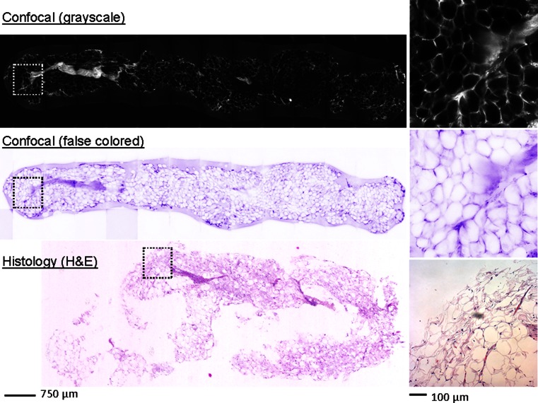
Tissue sampling is a problematic issue for inflammatory breast carcinoma, and immediate evaluation following core needle biopsy is needed to evaluate specimen adequacy. We sought to determine if confocal fluorescence microscopy provides sufficient resolution to evaluate specimen adequacy by comparing invasive tumor cellularity estimated from standard histologic images to invasive tumor cellularity estimated from confocal images of breast core needle biopsy specimens. Grayscale confocal fluorescence images of breast core needle biopsy specimens were acquired following proflavine application. A breast-dedicated pathologist evaluated invasive tumor cellularity in histologic images with hematoxylin and eosin staining and in grayscale and false-colored confocal images of cores. Agreement between cellularity estimates was quantified using a kappa coefficient. 23 cores from 23 patients with suspected inflammatory breast carcinoma were imaged. Confocal images were acquired in an average of less than 2 min per core. Invasive tumor cellularity estimated from histologic and grayscale confocal images showed moderate agreement by kappa coefficient: κ = 0.48 ± 0.09 (p < 0.001). Grayscale confocal images require less than 2 min for acquisition and allow for evaluation of invasive tumor cellularity in breast core needle biopsy specimens with moderate agreement to histologic images. We show that confocal fluorescence microscopy can be performed immediately following specimen acquisition and could indicate the need for additional biopsies at the initial visit.


Figures

Fig. 1
Fig. 1
Representative core with invasive tumor and non-neoplastic tissue (core 19); 25 % cellularity estimated by author S.K. in histologic image (standard H&E staining), 30 % cellularity estimated in grayscale confocal fluorescence image (0.01 % proflavine staining), and 25 % cellularity estimated in false-colored confocal fluorescence image derived from grayscale image (0.01 % proflavine staining). The solid outlined region indicates areas of the CNB specimen that contain invasive tumor. The scale bar at left is 750 μm. The insets at right show the border between neoplastic and non-neoplastic tissue in the CNB specimen. The locations of the insets are indicated in each core by a square with dashed lines. The scale bar at right is 100 μm
Fig. 2
Fig. 2
Representative core with invasive tumor (core 15); 80 % cellularity estimated by author S.K. in histologic image (standard H&E staining), 75 % cellularity estimated in grayscale confocal fluorescence image (0.01 % proflavine staining), and 90 % cellularity estimated in false-colored confocal fluorescence image derived from grayscale image (0.01 % proflavine staining). The solid outlined region indicates areas of the CNB specimen that contain neoplastic tissue. The scale bar at left is 750 μm. The insets at right show a region of neoplastic tissue in the CNB specimen. The locations of the insets are indicated in each core by a square with dashed lines. The scale bar at right is 100 μm
Fig. 3
Fig. 3
Representative core with no invasive tumor (core 22); 0 % cellularity estimated by author S.K. in histologic image (standard H&E staining), 0 % cellularity estimated in grayscale confocal fluorescence image (0.01 % proflavine staining), and 0 % cellularity estimated in false-colored confocal fluorescence image derived from grayscale image (0.01 % proflavine staining). This core is considered inadequate due to sampling error; no invasive tumor is visible despite a clinical indication of IBC. The scale bar at left is 750 μm. The insets at right show a region of non-neoplastic tissue in the CNB specimen. The locations of the insets are indicated in each core by a square with dashed lines. The scale bar at right is 100 μm
Fig. 4
Fig. 4
Scatterplots illustrating agreement on estimated invasive tumor cellularity between image types. Each data point represents a CNB specimen. Dashed lines represent the linear fit of the data. Pearson coefficients (R 2) quantify the linear fit of invasive tumor cellularity data. a Invasive tumor cellularity estimates from grayscale confocal images and histologic images. b Invasive tumor cellularity estimates from false-colored confocal images and histologic images

References

    1. Yamauchi H, Woodward WA, Valero V, Alvarez RH, Lucci A, Buchholz TA, et al. Inflammatory breast cancer: what we know and what we need to learn. Oncologist. 2012;17:891–899. doi: 10.1634/theoncologist.2012-0039. - DOI - PMC - PubMed
    1. Hance KW, Anderson WF, Devesa SS, Young HA, Levine PH. Trends in inflammatory breast carcinoma incidence and survival: the surveillance, epidemiology, and end results program at the National Cancer Institute. J Natl Cancer Inst. 2005;97:966–975. doi: 10.1093/jnci/dji172. - DOI - PMC - PubMed
    1. Anderson WF, Chu KC, Chang S. Inflammatory breast carcinoma and noninflammatory locally advanced breast carcinoma: distinct clinicopathologic entities? J Clin Oncol. 2003;21:2254–2259. doi: 10.1200/JCO.2003.07.082. - DOI - PubMed
    1. Dobbs JL, Ding H, Benveniste AP, Kuerer HM, Krishnamurthy S, Yang W, Richards-Kortum R. Feasibility of confocal fluorescence microscopy for real-time evaluation of neoplasia in fresh human breast tissue. J Biomed Opt. 2013 - PubMed
    1. Kumar N, Sayed S, Moloo Z, Wasike R. Fine-needle aspiration in suspected inflammatory breast cancer: case series with emphasis on approach to specimen adequacy. Acta Cytol. 2011;55:239–244. doi: 10.1159/000324032. - DOI - PubMed

Publication types