Review
. 2015 Jan;13(1):28-41.
doi: 10.1038/nrmicro3367. Epub 2014 Nov 24.

At the centre: influenza A virus ribonucleoproteins

Affiliations
Review

At the centre: influenza A virus ribonucleoproteins

Amie J Eisfeld et al. Nat Rev Microbiol. 2015 Jan.

Abstract

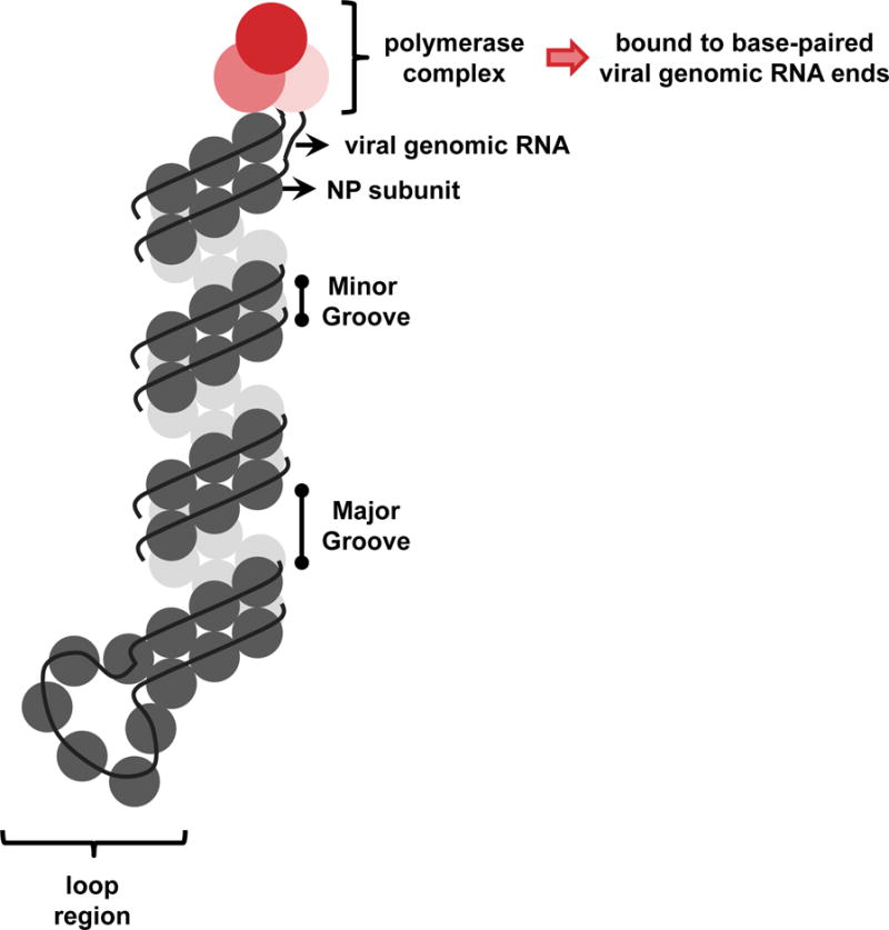
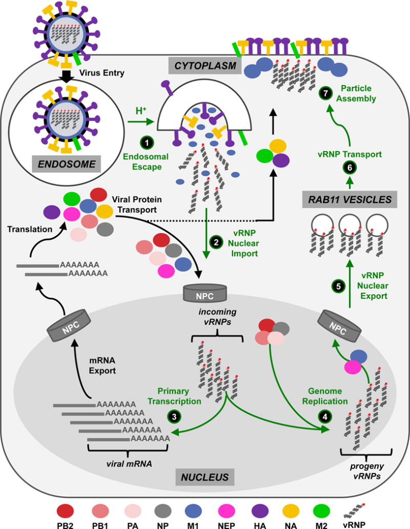
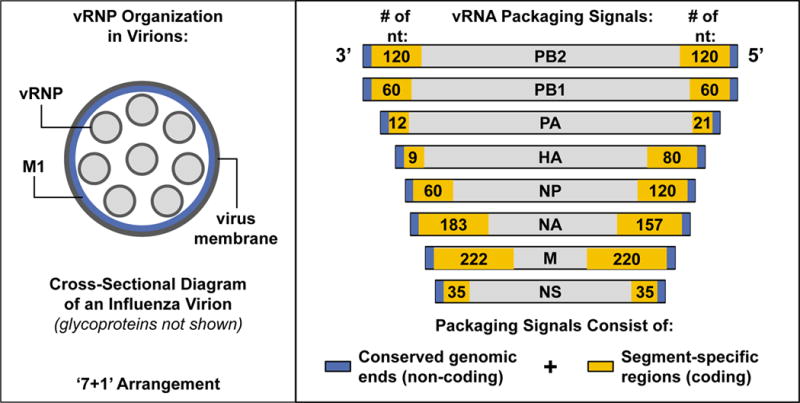
Influenza A viral ribonucleoprotein (vRNP) complexes comprise the eight genomic negative-sense RNAs, each of which is bound to multiple copies of the vRNP and a trimeric viral polymerase complex. The influenza virus life cycle centres on the vRNPs, which in turn rely on host cellular processes to carry out functions that are necessary for the successful completion of the virus life cycle. In this Review, we discuss our current knowledge about vRNP trafficking within host cells and the function of these complexes in the context of the virus life cycle, highlighting how structure contributes to function and the crucial interactions with host cell pathways, as well as on the information gaps that remain. An improved understanding of how vRNPs use host cell pathways is essential to identify mechanisms of virus pathogenicity, host adaptation and, ultimately, new targets for antiviral intervention.


Conflict of interest statement

COMPETING FINANCIAL INTERESTS

The authors declare that they have no competing financial interests.

Figures

Figure 1
Figure 1. Influenza vRNP complex
Each influenza vRNP consists of one single-stranded, negative-sense genomic RNA associated with multiple NP monomers and a single trimeric polymerase complex (composed of PB2, PB1, and PA). The 5′ and 3′ vRNA ends are complementary and base pair to form a double-stranded structure, which is bound by the polymerase complex at one end of the vRNP filament. The internal vRNA region is organized into an anti-parallel double helix, whose formation is driven by contacts between NP monomers (i.e., the ‘minor’ groove), and a loop can be observed at the end of the filament opposite to that bound by the polymerase complex.
Figure 2
Figure 2. Model for vRNP nuclear import
Uncoated, cytoplasmic vRNPs with exposed NP NLS motifs associate with IMPα, which in turn associates with IMPβ. The entire complex docks at the NPC and is transported into the nucleus, where Ran-GTP binds IMPβ and facilitates vRNP release into the nucleoplasm to initiate transcription and replication. Whether multiple IMPα and IMPβ molecules associate with each vRNP is unknown, as is the fate of the vRNP-associated IMPα once the vRNP cargo is released into the nucleoplasm.
Figure 3
Figure 3. Model for genome transcription and replication
(A, Primary Transcription) Initiation: Following nuclear entry, vRNPs that were associated with incoming viruses transcribe viral mRNAs in cis using the resident polymerase complex bound to the double-stranded genomic ends and cellular pre-mRNA caps obtained by cap-snatching from cellular RNA polymerase II. Elongation: Then, the vRNA is threaded through (and copied by) the viral polymerase complex. Poly-A Addition: When the 5′ end of the vRNA is reached, it is held by the polymerase to promote the generation of the poly-A tail in conjunction with the cellular SFPQ protein. (B, Genome Replication) After new viral proteins are translated by the cellular machinery, soluble polymerases mediate genome replication in trans, promoted by the activity of the cellular MCM complex and the Tat-SF1 and UAP56 cellular proteins. The FMR1 protein stimulates the assembly of polymerase complexes and NP in the presence of vRNA. The specific contributions of hCLE and IMPα (represented as ‘α’ in both the upper and lower panels) are currently unknown.
Figure 4
Figure 4. Model for vRNP nuclear export
Late in infection, vRNPs localize to the chromatin at the nuclear periphery and associate with components of the CRM1 nuclear export pathway (e.g., RCC1). After a signal that may include phosphorylation of an unknown target protein, mediated by signal transduction cascades activated by HA at the plasma membrane, the vRNPs are released from the chromatin. The vRNPs are subsequently exported from the nucleus by means of a ‘daisy chain’ complex, through which the viral NEP protein acts as an adaptor between vRNP-M1 and CRM1-Ran-GTP. The cellular YB-1 protein may be co-transported with vRNPs. At the cytoplasmic side of the NPC, Ran-GTP hydrolysis releases transport factors and vRNPs into the cytoplasm, where vRNPs remain associated with M1. Whether NEP remains associated with the cytoplasmic vRNPs remains unclear.
None
None

References

    1. WHO. Influenza (Seasonal) Fact sheet N°211. 2009 < http://www.who.int/mediacentre/factsheets/fs211/en/index.html>.
    1. Molinari NA, et al. The annual impact of seasonal influenza in the US: measuring disease burden and costs. Vaccine. 2007;25:5086–5096. doi: 10.1016/j.vaccine.2007.03.046. - DOI - PubMed
    1. Simonsen L. The global impact of influenza on morbidity and mortality. Vaccine. 1999;17(Suppl 1):S3–10. - PubMed
    1. van der Vries E, Schutten M, Fraaij P, Boucher C, Osterhaus A. Influenza virus resistance to antiviral therapy. Adv Pharmacol. 2013;67:217–246. doi: 10.1016/B978-0-12-405880-4.00006-8. - DOI - PubMed
    1. Matsuoka Y, et al. A comprehensive map of the influenza A virus replication cycle. BMC systems biology. 2013;7:97. doi: 10.1186/1752-0509-7-97. - DOI - PMC - PubMed

Publication types