. 2014 Dec;20(12):1999-2005.
doi: 10.3201/eid2012.141280.

Replication and shedding of MERS-CoV in upper respiratory tract of inoculated dromedary camels

Replication and shedding of MERS-CoV in upper respiratory tract of inoculated dromedary camels

Danielle R Adney et al. Emerg Infect Dis. 2014 Dec.

Abstract

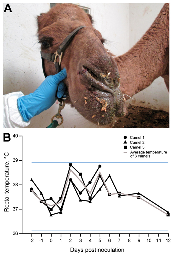
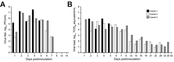
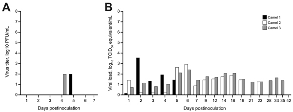
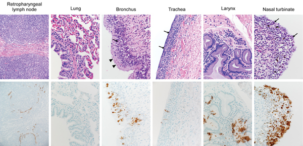
In 2012, a novel coronavirus associated with severe respiratory disease in humans emerged in the Middle East. Epidemiologic investigations identified dromedary camels as the likely source of zoonotic transmission of Middle East respiratory syndrome coronavirus (MERS-CoV). Here we provide experimental support for camels as a reservoir for MERS-CoV. We inoculated 3 adult camels with a human isolate of MERS-CoV and a transient, primarily upper respiratory tract infection developed in each of the 3 animals. Clinical signs of the MERS-CoV infection were benign, but each of the camels shed large quantities of virus from the upper respiratory tract. We detected infectious virus in nasal secretions through 7 days postinoculation, and viral RNA up to 35 days postinoculation. The pattern of shedding and propensity for the upper respiratory tract infection in dromedary camels may help explain the lack of systemic illness among naturally infected camels and the means of efficient camel-to-camel and camel-to-human transmission.


Figures

Figure 1
Figure 1
Clinical signs in dromedary camels inoculated with Middle East respiratory syndrome coronavirus (MERS-CoV). A) Nasal discharge observed in camel 3; each of 3 inoculated camels had nasal discharge during the first 2 weeks of the experiment. B) Rectal temperatures are indicated for each camel by lines with geometric shapes. Horizontal lines indicate the normal temperature range observed among these dromedary camels as calculated by mean ± 3×, the before inoculation.
Figure 2
Figure 2
Virus shedding from the upper respiratory tract in dromedary camels inoculated Middle East respiratory syndrome coronavirus (MERS-CoV). Shedding was determined by A) infectious titers by plaque assay and B) viral load by quantitative real-time PCR. We extrapolated 50% tissue culture infective dose (TCID50) equivalents from standard curves generated by 10-fold dilutions of a MERS-CoV stock (HCoV-EMC/2012) with known virus titer in parallel to each quantitative real-time PCR run.
Figure 3
Figure 3
Virus shedding determined by oral swabs obtained from dromedary camels inoculated with Middle East respiratory syndrome coronavirus (MERS-CoV). Shedding was determined by A) determining infectious titers and B) viral RNA. Virus titers were determined by plaque assay and viral load by qRT-PCR. TCID50 equivalents were extrapolated from standard curves generated by 10-fold dilutions of a MERS-CoV stock (HCoV-EMC/2012) with known virus titer in parallel to each run.
Figure 4
Figure 4
Virus titers in tissues collected from dromedary camel 1 inoculated with Middle East respiratory syndrome coronavirus (MERS-CoV). Tissues were collected at 5 days postinoculation (dpi) for camel 1, 28 dpi for vamel 2 and 42 dpi for camel 3. Detectable infectious virus in the collected tissues was found only in camel 1. Nasal turbinates were sampled in 3 different sections: anterior, medial, and posterior. Infectious titers were determined by plaque assay. LN, lymph node.
Figure 5
Figure 5
Histopathologic changes at 5 days postinoculation in camel 1 inoculated with Middle East respiratory syndrome coronavirus (MERS-CoV). Tissues were collected and stained with hematoxylin and eosin (top row). Anti–MERS-CoV immunohistochemical results (bottom row) are visible as a red-brown stain. Degeneration of the pseudostratified epithelium lining the nasal turbinate, trachea, and bronchus is indicated by the absence of goblet cells, cilia and nuclear regimentation with infiltration of neutrophils (arrows). The arrowheads indicate areas where the cilia remained intact. Original magnification ×400.

References

    1. Zaki AM, van Boheemen S, Bestebroer TM, Osterhaus AD, Fouchier RA. Isolation of a novel coronavirus from a man with pneumonia in Saudi Arabia. N Engl J Med. 2012;367:1814–20. 10.1056/NEJMoa1211721 - DOI - PubMed
    1. World Health Organization. Global Alert and Response. Middle East respiratory syndrome coronavirus (MERS-CoV) – update; 2014. June [cited 30 Jun 2014]. http://www.who.int/csr/don/2014_06_16_mers/en/
    1. Puzelli S, Azzi A, Santini MG, Di Martino A, Facchini M, Castrucci MR, et al. Investigation of an imported case of Middle East respiratory syndrome coronavirus (MERS-CoV) infection in Florence, Italy, May to June 2013. Euro Surveill. 2013;18:20564 . - PubMed
    1. Milne-Price S, Miazgowicz KL, Munster VJ. The emergence of the Middle East respiratory syndrome coronavirus. Pathog Dis. 2014;71:121–36. - PMC - PubMed
    1. Assiri A, McGeer A, Perl TM, Price CS, Al Rabeeah AA, Cummings DA, et al. Hospital outbreak of Middle East respiratory syndrome coronavirus. N Engl J Med. 2013;369:407–16. 10.1056/NEJMoa1306742 - DOI - PMC - PubMed

Publication types

MeSH terms