. 2014 Dec;20(12):2006-14.
doi: 10.3201/eid2012.140548.

Transmission characteristics of variably protease-sensitive prionopathy

Transmission characteristics of variably protease-sensitive prionopathy

Silvio Notari et al. Emerg Infect Dis. 2014 Dec.

Abstract

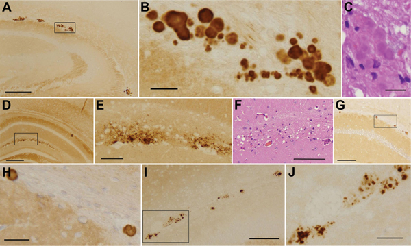
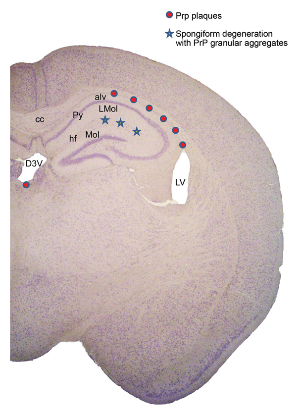
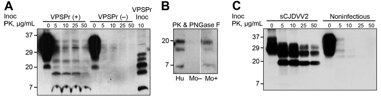
Variably protease-sensitive prionopathy (VPSPr), a recently identified and seemingly sporadic human prion disease, is distinct from Creutzfeldt-Jakob disease (CJD) but shares features of Gerstmann-Sträussler-Scheinker disease (GSS). However, contrary to exclusively inherited GSS, no prion protein (PrP) gene variations have been detected in VPSPr, suggesting that VPSPr might be the long-sought sporadic form of GSS. The VPSPr atypical features raised the issue of transmissibility, a prototypical property of prion diseases. We inoculated VPSPr brain homogenate into transgenic mice expressing various levels of human PrP (PrPC). On first passage, 54% of challenged mice showed histopathologic lesions, and 34% harbored abnormal PrP similar to that of VPSPr. Surprisingly, no prion disease was detected on second passage. We concluded that VPSPr is transmissible; thus, it is an authentic prion disease. However, we speculate that normal human PrPC is not an efficient conversion substrate (or mouse brain not a favorable environment) and therefore cannot sustain replication beyond the first passage.


Figures

Figure 1
Figure 1
Histologic and immunohistochemical findings from a study of the transmission characteristics of VPSPr. A‒E). Two types of lesions typically observed in Tg(HuPrP129V)×8 mice. A‒C) Plaques are most often located at the border between the alveus of the hippocampus and the corpus callosum, often forming aggregates distributed in a row. They can be tightly aggregated or partially fused generating multicore plaques. PrP immunostaining (A, B) shows well-formed plaques surrounded by PrP deposits that appear in various stages of aggregation at HE. C), where plaque cores appear smooth, lacking the spiny appearance of typical kuru plaques. D‒F) The lesion of the second type consists of PrP granular deposits (D and E) co-localized with spongiform degeneration in the layer lacunosum moleculare of the hippocampus (F). G‒J) Plaques in Tg(HuPrP129V)×3 mice were fewer but similar in location and appearance to those of Tg(HuPrP129V)×8. In contrast, PrP aggregates generally appeared to be loose and formed fewer real plaques in Tg(HuPrP129M)×2–positive mice (I and J). PrP monoclonal antibody 3F4. The boxes in panels A, D, G, and I mark the exact areas that are shown at higher magnification in panels B, E, H, and J, respectively. PrP, prion protein; VPSPr, variably protease-sensitive prionopathy; Tg, transgenic. Scale bars in A and E = 250 μm. Scales bar in B, C, H, and J = 25 μm. Scale bar in D = 500 μm. Scale bars in F, G, and I = 100 μm.
Figure 2
Figure 2
Representation of lesion topography in brain of positive Tg(HuPrP129V)×8 mice inoculated with VPSPr-129VV brain homogenate. alv, alveus of hippocampus; cc, corpus callosum; D3V, dorsal third ventricle; hf, hippocampal fissure; L mol, lacunosum molecular layer; LV, lateral ventricle; Mol, molecular layer dentate gyrus; Py, pyramidal cell layer of hippocampus.
Figure 3
Figure 3
Western blot characteristics of PrPSc recovered from brain of VPSPr-inoculated Tg mice and controls. A) BH treated with increasing amounts of PK show PK-resistant PrPSc fragments with a ladder-like electrophoretic profile in positive VPSPr-inoculated mice, VPSPr (+), even at high concentrations of PK (50 μL/mL). In contrast, nonspecific bands are seen in negative VPSPr-inoculated mice, VPSPr (‒). The banding pattern in VPSPr (+) roughly recapitulates that of the PK-treated PrPSc from the VPSPr inoculum (VPSPr Inoc). B) Positive Tg mice BH treated with PK (25 μg/mL) and PNGase F show 3 PrPSc bands migrating at ≈20 kDa, ≈17 kDa, and ≈7 kDa (Mo +), replicating those of similarly treated VPSPr inoculum (Hu). No bands can be detected in the negative Tg mice (Mo ‒). (All 3 preparations were run on the same gel, but unnecessary lanes were removed). C) Tg mice inoculated with sCJDVV2 BH (from sCJD homozygous valine harboring PrPSc type 2) or inoculated with noninfectious BH used as positive and negative controls exhibit typical PK-resistant PrPSc. Tg(HuPrP129V)×8 BH and monoclonal antibody 1E4 were used in all Western blot tests. Approximate molecular masses are in kDa. BH, brain homogenate; PK, proteinase K; PrPSc, scrapie prion protein; sCJD, sporadic Creutzfeldt-Jakob disease; VPSPr, variably protease-sensitive prionopathy; Tg, transgenic.

References

    1. Prusiner SB. Biology and genetics of prions causing neurodegeneration. Annu Rev Genet. 2013;47:601–3. 10.1146/annurev-genet-110711-155524 - DOI - PMC - PubMed
    1. Surewicz WK, Apostol MI. Prion protein and its conformational conversion: a structural perspective. Top Curr Chem. 2011;305:135–67. 10.1007/128_2011_165 - DOI - PubMed
    1. Aguzzi A, Falsig J. Prion propagation, toxicity and degradation. Nat Neurosci. 2012;15:936–9. 10.1038/nn.3120 - DOI - PubMed
    1. Sandberg MK, Al-Doujaily H, Sharps B, Clarke AR, Collinge J. Prion propagation and toxicity in vivo occur in two distinct mechanistic phases. Nature. 2011;470:540–2. 10.1038/nature09768 - DOI - PubMed
    1. Gambetti P, Cali I, Notari S, Kong Q, Zou WQ, Surewicz WK. Molecular biology and pathology of prion strains in sporadic human prion diseases. Acta Neuropathol. 2011;121:79–90. 10.1007/s00401-010-0761-3 - DOI - PMC - PubMed

Publication types