. 2015 Oct;33(10):1455-65.
doi: 10.1002/jor.22791. Epub 2015 Aug 12.

Autologous implantation of BMP2-expressing dermal fibroblasts to improve bone mineral density and architecture in rabbit long bones

Affiliations

Autologous implantation of BMP2-expressing dermal fibroblasts to improve bone mineral density and architecture in rabbit long bones

Akikazu Ishihara et al. J Orthop Res. 2015 Oct.

Abstract

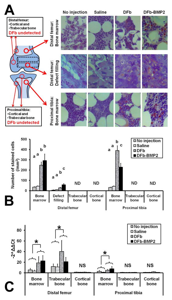
Cell-mediated gene therapy may treat bone fragility disorders. Dermal fibroblasts (DFb) may be an alternative cell source to stem cells for orthopedic gene therapy because of their rapid cell yield and excellent plasticity with bone morphogenetic protein-2 (BMP2) gene transduction. Autologous DFb or BMP2-expressing autologous DFb were administered in twelve rabbits by two delivery routes; a transcortical intra-medullar infusion into tibiae and delayed intra-osseous injection into femoral drill defects. Both delivery methods of DFb-BMP2 resulted in a successful cell engraftment, increased bone volume, bone mineral density, improved trabecular bone microarchitecture, greater bone defect filling, external callus formation, and trabecular surface area, compared to non-transduced DFb or no cells. Cell engraftment within trabecular bone and bone marrow tissue was most efficiently achieved by intra-osseous injection of DFb-BMP2. Our results suggested that BMP2-expressing autologous DFb have enhanced efficiency of engraftment in target bones resulting in a measurable biologic response by the bone of improved bone mineral density and bone microarchitecture. These results support that autologous implantation of DFb-BMP2 warrants further study on animal models of bone fragility disorders, such as osteogenesis imperfecta and osteoporosis to potentially enhance bone quality, particularly along with other gene modification of these diseases.

Keywords: autologous implantation; bone morphogenetic protein-2; dermal fibroblasts; intra-medullar cell delivery; intra-osseous cell delivery.


Conflict of interest statement

All authors have no conflict of interest to declare.

Figures

Figure 1
Figure 1
Experimental models (A), protocol (B), and assignment table (C) of the in vivo studies. Twelve rabbits received intra-medullar infusion and intra-osseous infusion of autologous dermal fibroblasts (DFb) transduced by FV vector carrying green florescent protein (GFP) genes, subsequently non-transduced or transduced by Adenoviral (Ad) vectors encoding morphogenetic protein-2 (BMP2) genes, as illustrated. *One of 12 rabbits (DFb-BMP2 injection/infusion in one hindlimb and saline injection/infusion in the contralateral limb) died at day 2 with unknown reason and was not included in any outcome assessments.
Figure 2
Figure 2
Gene transduction by Adenoviral (Ad) vector carrying green fluorescent protein (GFP) genes was significantly greater (*P<0.02) in MSC compared to dermal fibroblasts (DFb) at 50, 100, and 150 moi (multiplicities of infection), but it was comparable between MSC and DFb at 200 moi (A; left), and the bone morphogenetic protein-2 (BMP2) production measured by ELISA was significantly greater (#P<0.04) in MSC with 100 or 200 moi Ad-BMP2 or DFb with 200 moi Ad-BMP2 compared to DFb with 100 moi Ad-BMP2 (A; right). When 200 moi Ad-BMP2 was applied, DFb and MSC showed comparable level of mineralized nodule formation (Von Kossa staining) at days 7 and 14 (B). The cell-sorting procedures of DFb with FV-GFP transduction using flow-cytometry were facilitated to increase the proportion of GFP labeled cells (C; left), and the GFP gene expression sustained similar level up to 28 days after the cell-sortings (C; right). Data were expressed as mean+s.e.m. NS: there were no significant differences among treatment groups.
Figure 3
Figure 3
Micro-computed tomographic (μCT) images (A) and quantitative analysis of efficacy of dermal fibroblast (DFb)-mediated bone morphogenetic protein-2 (BMP2) gene therapy in limbs ofrabbits after intra-osseous injection and intra-medullar infusion of DFb or DFb-BMP2 (Fig 1B, 1D, and 1E). In cortical bone regions of distal femur and proximal tibiae, bone volume (B; left) and density (B; right) were not different among treatments. In trabecular bone regions of distal femur and proximal tibiae, bone volume (C; upper left), bone density (C; upper right), bone surface area (C; lower left), and trabecular thickness (C; lower right) were significantly greater (P<0.03) in DF-BMP2 injected/infused bone tissues compared to other 3 groups. In external callus regions of distal femur, bone volume (D; left) was significantly greater (P<0.02) in DF-BMP2 injected/infused bone tissues compared to other 3 groups, although bone density (D; right) was not different among treatments. In drill defect regions of distal femur, bone volume (E; left) was significantly greater (P<0.01) in DF-BMP2 injected/infused bone tissue compared to the other 3 groups, although bone density (E; right) was not different among treatments. Data were expressed as mean+s.e.m. abcDifferent letters differ significantly (P<0.05). NS: there were no significant differences among treatment groups.
Figure 4
Figure 4
Histomorphometric evaluation of the distal femurs (A; left) and proximal tibiae (A; right) for the efficacy of dermal fibroblast (DFb)-mediated bone morphogenetic protein-2 (BMP2) gene therapy in limbs of rabbits after intra-osseous injection and intra-medullar infusion of DFb or DFb-BMP2 (Fig 1B, 1D, and 1E). In cortical bone regions of distal femur and proximal tibiae, epiphyseal cortical thickness (B; left) and subchondral bone thickness (B; right) were not different among treatments. In trabecular bone regions of distal femur and proximal tibiae, bone area (C; upper left), fractal dimension (C; upper right), inter-connectivity index (C; lower left), and Euler number (C; lower right) were significantly greater (P<0.04) in DF-BMP2 injected/infused bone tissues compared to other 3 groups. In drill defect regions of distal femur, porosity (D; left) was significantly less (P<0.007) and maturity (D; right) was significantly greater (P<0.02) in DF-BMP2 injected/infused bone tissues compared to other 3 groups. Data were expressed as mean+s.e.m. abcDifferent letters differ significantly (P<0.05). NS: there were no significant differences among treatment groups.
Figure 5
Figure 5
Immunohistochemical (IHC) evaluation (A,B) and RT-PCR analysis (C) for the efficacy of dermal fibroblast (DFb)-mediated bone morphogenetic protein-2 (BMP2) gene therapy in limbs of rabbits after intra-osseous injection and intra-medullar infusion of DFb or DFb-BMP2 (Fig 1B, 1D, and 1E). In the bone marrow or defect filling tissues of distal femurs, the number of GFP-positive cells was significantly greater (P<0.003) in DFb or DF-BMP2 injected/infused bone tissues compared to no-injection or saline groups. In the bone marrow tissues of proximal tibiae, the number of GFP-positive cells was significantly greater (P<0.001) in DFb or DF-BMP2 injected/infused bone tissues compared to no-injection or saline groups. In the bone marrow or trabecular bone tissues of distal femurs, the GFP-gene expression was significantly greater (P<0.04) in DFb and DF-BMP2 injected/infused bone tissues compared to no-injection and saline groups. In the bone marrow tissues of proximal tibiae, the GFP-gene expression was significantly greater (P<0.04) in DFb and DF-BMP2 injected/infused bone tissues compared to no-injection and saline groups.

Similar articles

Cited by

References

    1. Rauch F, Glorieux FH. Osteogenesis imperfecta. Lancet. 2004;363:1377–1385. - PubMed
    1. Rossini M, Di Munno O, Gatti D, et al. Optimising bisphosphonate treatment outcomes in postmenopausal osteoporosis: review and Italian experience. Clin Exp Rheumatol. 2011;29:728–735. - PubMed
    1. Munns CF, Rauch F, Mier RJ, et al. Respiratory distress with pamidronate treatment in infants with severe osteogenesis imperfect. Bone. 2004;35:231–234. - PubMed
    1. Pizones J, Plotkin H, Parra-Garcia JI, et al. Bone healing in children with osteogenesis imperfecta treated with bisphosphonates. J Pediatr Orthop. 2005;25:332–335. - PubMed
    1. Chan B, Zacharin M. Maternal and infant outcome after pamidronate treatment of polyostotic fibrous dysplasia and osteogenesis imperfecta before conception: a report of four cases. J Clin Endocrinol Metab. 2006;91:2017–2020. - PubMed

Publication types

Substances