. 2014 Oct 15;7(10):3181-92.
eCollection 2014.

Increased circulating myeloid-derived suppressor cells correlate with cancer stages, interleukin-8 and -6 in prostate cancer

Affiliations

Increased circulating myeloid-derived suppressor cells correlate with cancer stages, interleukin-8 and -6 in prostate cancer

Ning Chi et al. Int J Clin Exp Med. .

Abstract

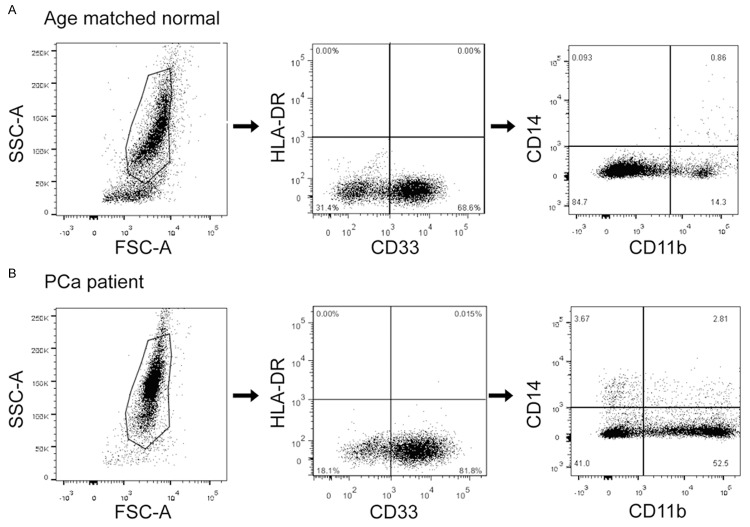
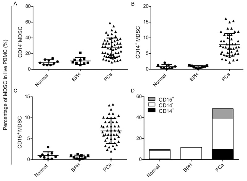
Aim: Myeloid-derived suppressor cells (MDSCs) are a population of cells which negatively regulate immune response during tumor progression. In this study, we assessed the accumulation of MDSCs (CD33(+)CD11b(+)HLA-DR(-)CD14(-)) in patients with prostate cancer and its clinical relevance.

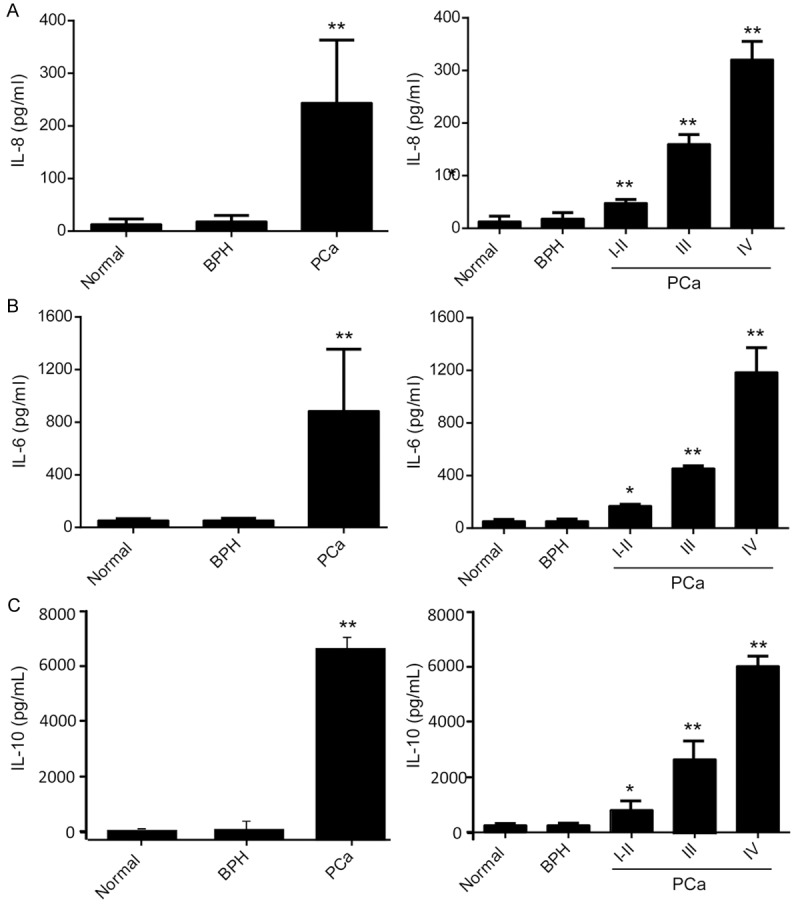
Methods: We tested the frequency of MDSCs in the peripheral blood of patients with prostate cancer or benign prostate hyperplasia and healthy donors. Serumal interleukin-8, -6 and -10 were analyzed. Effects of MDSCs on the T cell response were determined.

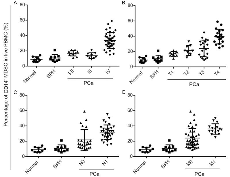
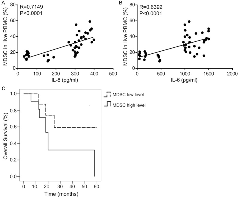
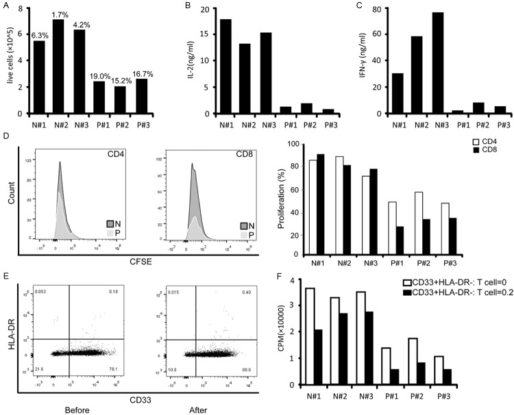
Results: MDSCs increased in cancer patients, and there was an association between MDSCs and cancer stages or overall survival. Elevated serumal interleukin-8 and -6 in cancer patients correlated with MDSCs. Moreover, accumulation of MDSCs was associated with defective T cell function.

Conclusion: Our study showed an increased population of MDSCs in patients with prostate cancer. Interleukin-8 and -6 in serum may play a new important role companied with MDSCs in prostate cancer.

Keywords: IL-6; IL-8; MDSCs; prostate cancer.


Figures

Figure 1
Figure 1
MDSCs FACS and scatter plots of cancer patients and healthy controls. CD33+CD11b+HLA-DR-CD14- MDSCs FACS gating of PBMC of a patient with prostate cancer (A) and an age-matched normal control (B). Dot plots represent live gated events. The forward and side scatter gate was analyzed for CD33+HLA-DR- cells. Then the CD33+HLA-DR- gate was analyzed for cells expressing CD11b and CD14. MDSCs were calculated as a percentage of live cells in PBMCs. Markers analyzed are indicated in the axis of each FACS plot.
Figure 2
Figure 2
Significantly elevated MDSCs in patients with prostate cancer. A. Mean granulocytic CD33+CD11b+HLA-DR-CD14- MDSCs levels in patients with prostate cancer were significantly higher than age-matched controls and patients with BPH. B. Mean monocytic CD33+CD11b+HLA-DR-CD14+ MDSCs levels in patients with prostate cancer were higher than controls and patients with BPH. C. Mean granulocytic CD33+CD11b+HLA-DR-CD15+ MDSCs levels in patients with prostate cancer were higher than controls and patients with BPH. D. CD33+CD11b+HLA-DR-CD14- MDSCs is the the major phenotype of MDSCs in prostate cancer.
Figure 3
Figure 3
CD33+CD11b+HLA-DR-CD14- MDSCs correlated with clinical stages. Correlation between percentage of MDSCs and clinical stages (A), T classification (B), N classification (C) and distant metastasis (D) in patients with prostate cancer and normal controls (*P<0.05, **P<0.01).
Figure 4
Figure 4
IL-8, -6 and -10 are significantly higher expressed in patients with prostate cancer and correlated with stages of cancer. Serum cytokines levels including IL-8 (A), IL-6 (B) and IL-10 (C) in age-matched normal controls and patients with prostate cancer were measured using ELISA assays. Serum samples from patients had a significantly higher level of IL-8, 6 and 10 than control samples (left lanes) and the levels increased with clinical stages (right lanes) (*P<0.05, **P<0.01).
Figure 5
Figure 5
MDSCs correlated with serum IL-8 and IL-6 in patients with prostate cancer. MDSCs were associated with the poor prognosis of patients with prostate cancer. Spearman correlation analyses between percentages of MDSCs and serum levels of IL-8 (A, r=0.7149, P<0.0001) and IL-6 (B, r=0.6392, P<0.0001) in patients with prostate cancer. (C) Correlation between percentages of MDSCs and survival by Kaplan-Meier analysis of patients with the high (≥ the median) or low (< the median) MDSCs level.
Figure 6
Figure 6
MDSCs decreased T cell responses. Equal numbers of PBMCs from normal controls (N) and three patients with stage IV cancer (P) were assayed for cell proliferation (A), IL-2 (B) and IFN-γ(C) secretion in response to activation with anti-CD3/CD28-coated beads. Corresponding percentages of circulating MDSCs are shown above in bold. D. PBMCs were stained with CFSE, cultured for 5 days, and then stained with monoclonal antibodies against CD4 and CD8; proliferation was quantified as the percentages of CFSElow cells. Left: Representative histogram of the FACS analysis. Right: Analysis of T cell proliferation. Myeloid cells (CD33+HLA-DR-) were isolated from freshly drawn blood from three normal controls (N) and three patients with stage IV cancer (P). Direct contact of T cells with isolated myeloid cells (CD33+HLA-DR-) from cancer patients inactivates T cell. E. Representative histograms of CD33+HLA-DR- fractions before and after enrichment. F. Proliferation of isolated autologous T cells in response to CD3/CD28 activation in the presence of the indicated ratios of purified autologous CD33+HLA-DR- cells.

References

    1. Ochando JC, Chen SH. Myeloid-derived suppressor cells in transplantation and cancer. Immunol Res. 2012;54:275–285. - PMC - PubMed
    1. Brusa D, Simone M, Gontero P, Spadi R, Racca P, Micari J, Degiuli M, Carletto S, Tizzani A, Matera L. Circulating immunosuppressive cells of prostate cancer patients before and after radical prostatectomy: profile comparison. Int J Urol. 2013;20:971–978. - PubMed
    1. Marigo I, Dolcetti L, Serafini P, Ostrand-Rosenberg S. Tumor-induced tolerance and immune suppression by myeloid derived suppressor cells. Immunol Rev. 2008;222:162–179. - PubMed
    1. Kong YY, Fuchsberger M, Xiang SD, Apostolopoulos V, Plebanski M. Myeloid derived suppressor cells and their role in diseases. Curr Med Chem. 2013;20:1437–1444. - PubMed
    1. Diaz-Montero CM, Salem ML, Nishimura MI, Garrett-Mayer E, Cole DJ, Montero AJ. Increased circulating myeloid-derived suppressor cells correlate with clinical cancer stage, metastatic tumor burden, and doxorubicin-cyclophosphamide chemotherapy. Cancer Immunol Immunother. 2009;58:49–59. - PMC - PubMed

LinkOut - more resources