. 2015 Jan;16(1):67-74.
doi: 10.1038/ni.3046. Epub 2014 Nov 24.

The methyltransferase Setdb2 mediates virus-induced susceptibility to bacterial superinfection

Affiliations

The methyltransferase Setdb2 mediates virus-induced susceptibility to bacterial superinfection

Christopher Schliehe et al. Nat Immunol. 2015 Jan.

Abstract

Immune responses are tightly regulated to ensure efficient pathogen clearance while avoiding tissue damage. Here we report that Setdb2 was the only protein lysine methyltransferase induced during infection with influenza virus. Setdb2 expression depended on signaling via type I interferons, and Setdb2 repressed expression of the gene encoding the neutrophil attractant CXCL1 and other genes that are targets of the transcription factor NF-κB. This coincided with occupancy by Setdb2 at the Cxcl1 promoter, which in the absence of Setdb2 displayed diminished trimethylation of histone H3 Lys9 (H3K9me3). Mice with a hypomorphic gene-trap construct of Setdb2 exhibited increased infiltration of neutrophils during sterile lung inflammation and were less sensitive to bacterial superinfection after infection with influenza virus. This suggested that a Setdb2-mediated regulatory crosstalk between the type I interferons and NF-κB pathways represents an important mechanism for virus-induced susceptibility to bacterial superinfection.


Figures

Figure 1
Figure 1
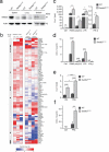
Setdb2 is induced upon influenza virus infection and TLR stimulation in an IFNAR1 dependent manner. (a) WT mice were intranasally infected with influenza virus or mock treated. 18 hours later lung sections were stained using an H1N1-specific antibody. H1N1 infected areas are indicated by arrowheads. Scale bar, 250μm. Representative images for 3 individual mice are shown. (b) Expression profile of protein lysine methyltransferases (PKMTs) in lungs upon influenza virus infection (PR8) compared to uninfected controls (UI) (n=6, 2 experiments independent pooled). Heatmap illustrations of (left) log2-fold change compared to uninfected mice respectively (right) Robust Multi-array Average (RMA) values are shown. (c) WT, Ifnar1–/–, Irf7–/– and Stat1–/– mice were intranasally infected with influenza virus (PR8) or mock treated (Ctrl). 18 hours later lung tissue was collected and expression of Setdb2 was determined by real-time PCR. Scatter blots represent the fold change of Setdb2 expression of individual mice compared to uninfected WT mice. Data points were pooled from 2 independent experiments. (d-f) BMDMs from Ifnar–/– and WT mice were either left untreated (Ctrl), (d) infected with influenza virus (PR8, MOI 10), (e) stimulated with IFN-β, IFN-γ or IFN-λ, or (f) stimulated with the indicated TLR agonists PAM3, poly(I:C), and LPS. Setdb2 mRNA expression was quantified by real-time PCR 8 hours after stimulation and is displayed as fold induction compared to untreated WT cells. Biological triplicates of one out of two similar independent experiments are shown. For protein detection, Setdb2 expression was analyzed (d) 8 hours respectively (e, f) 24 hours after stimulation by immunoblot using the Setdb2-specific mAb clone 7H7F11. Antibodies specific for the IFN-stimulated protein Zbp1 and actin served as controls for induction and loading, respectively. (d, e) Immunoblots show results of one out of two representative experiments. (c-f) Statistical significance was calculated by unpaired t-test. Significant p-values were indicated as follows: * p≤0.05, ** p≤0.01, *** p≤0.001, **** p≤0.0001.
Figure 2
Figure 2
Setdb2GT/GT macrophages show increased expression of a subset of NF-κB target genes including CXCL1. (a) Spleen and lung tissue as well as bone marrow-derived macrophages (BMDMs) from naive Setdb2GT/GT or WT mice were analyzed for the expression of Setdb2 by immunoblot using the Setdb2-specific mAb clone 7H7F11. Actin served as loading control. (b) BMDMs were treated with poly(I:C) for 0, 2 and 8 hours and expression profiling was performed by RNAseq using biological triplicates for each condition. Heatmap illustrations for (left) differential gene expression between WT and Setdb2GT/GT BMDMs of all protein-coding genes, respectively (right) the expression profiles at 2 and 8 hours compared to untreated BMDMs of the respective genotype are shown. NF-κB target genes are indicated as grey boxes. BMDMs from either WT or Setdb2GT/GT mice were either left untreated (Ctrl), (c, d) stimulated with the indicated TLR agonists and with IFN-β or (e, f) infected with influenza virus (PR8, MOI 10). (c, e) Cxcl1 mRNA expression was quantified by real-time PCR after 2 hours of stimulation. (d, f) CXCL1 secretion was quantified by ELISA after 8 hours of stimulation. Biological triplicates of one out of two similar independent experiments are shown. (c-f) Statistical significance was calculated by unpaired t-test. Significant p-values were indicated as follows: * p≤0.05, ** p≤0.01, *** p≤0.001, **** p≤0.0001.
Figure 3
Figure 3
Setdb2 binds to the Cxcl1 promoter region and associates with H3K9 trimethylation. (a-c) WT or Setdb2GT/GT BMDMs were stimulated with poly(I:C) for 2 hours or left untreated (Ctrl) before cells were prepared for chromatin immunoprecipitation (ChIP). Specific enrichment of indicated promoter elements, the Cxcl1 promoter (prom) and the Cxcl1 exon 1 (ex1), was quantified by real-time PCR. Data are depicted as normalized recovery to the highest value in WT of duplicate measurements from two independent experiments (a, b), and one experiment (c). Empty beads from one respective experiment were used as mock (M). (a) ChIP for endogenous Setdb2 using the Setdb2-specific mAb clone 7H7F11. (b) ChIP for H3K9me3. (c) ChIP for H3K9ac. Statistical significance was calculated by unpaired t-test. Significant p-values were indicated as follows: * p≤0.05, ** p≤0.01, *** p≤0.001, **** p≤0.0001.
Figure 4
Figure 4
Setdb2GT/GT mice show exacerbated lung inflammation in a model of LPS-induced neutrophilia. WT and Setdb2GT/GT mice were intranasally challenged with LPS for 4 hours or mock treated (Ctrl). (a) Total lung RNA from challenged and control mice was extracted and analyzed for the expression of Cxcl1 by real-time PCR. (b) Bronchoalveolar lavage (BAL) was performed and BAL fluid supernatants were analyzed for CXCL1 by ELISA. (c-e), Total BAL fluid cells as well as neutrophils and macrophages were quantified. Cell numbers of individual experiments were normalized to the respective means of LPS-stimulated WT mice (relative number = cell number/meanWT+LPS). Scatter blots represent individual mice pooled from 3 independent experiments. Statistical analysis was performed by unpaired t-test. Significant p-values were indicated as follows: * p≤0.05, ** p≤0.01, *** p≤0.001, **** p≤0.0001.
Figure 5
Figure 5
Setdb2 mediates influenza virus-induced susceptibility to superinfection with Streptococcus pneumoniae. WT and Setdb2GT/GT mice were either left untreated (Ctrl), infected with a sublethal dose of influenza virus (PR8) or superinfected with Streptococcus pneumoniae (SP) 5 days after PR8 infection. (a) Cxcl1 RNA was quantified by real-time PCR from lung tissue 5 days after influenza virus infection. (b-d) WT and Setdb2GT/GT mice were harvested 16 hours after SP superinfection. Superinfected mice received ~2×10⁴ CFU of SP. (b) Results of CXCL1 ELISA from BAL fluid and enumeration of neutrophils from (c) lung tissue and (d) BAL fluid are shown. (e-k) 2 days after superinfection with ~2×10³ CFU of SP, WT and Setdb2GT/GT mice were sacrificed and (e) images of lungs were taken (size bar, 1cm). Lungs were aligned according to the degree of gross pathology. (f) The lung wet weight was determined from the right lobes and normalized to the body weight on day zero. A dotted line depicts the average lung weight to body weight ratio of three uninfected lungs. (g) H&E histological staining of representative sections of the left lung lobes of WT and Setdb2GT/GT mice are shown. Scale bar of low magnification is 200μm, high magnification 50μm. (h) Histopathological scoring of lung sections (see Methods). (i) Expression of Il6 mRNA in lung tissue was determined by real-time PCR. Graph shows Il6 fold induction compared to uninfected WT mice. (j) Levels of IL-6 protein were detected by ELISA. (k) Bacterial burden was determined as CFU in tissue homogenates of the right lung lobes. Scatter blots represent individual mice from (a, e-k) one or (b-d) two pooled experiments. (f, k) show one out of two similar experiments. Statistical significance was calculated by unpaired t-test. Significant p-values were indicated as follows: * p≤0.05, ** p≤0.01, *** p≤0.001, **** p≤0.0001.

Comment in

  • Stop the executioners.
    Wack A. Wack A. Nat Immunol. 2015 Jan;16(1):6-8. doi: 10.1038/ni.3055. Nat Immunol. 2015. PMID: 25521671 No abstract available.

References

    1. Morens DM, Taubenberger JK, Fauci AS. Predominant role of bacterial pneumonia as a cause of death in pandemic influenza: implications for pandemic influenza preparedness. The Journal of infectious diseases. 2008;198(7):962–970. - PMC - PubMed
    1. Molinari NA, Ortega-Sanchez IR, Messonnier ML, Thompson WW, Wortley PM, Weintraub E, et al. The annual impact of seasonal influenza in the US: measuring disease burden and costs. Vaccine. 2007;25(27):5086–5096. - PubMed
    1. van der Sluijs KF, van der Poll T, Lutter R, Juffermans NP, Schultz MJ. Bench-to-bedside review: bacterial pneumonia with influenza - pathogenesis and clinical implications. Critical care. 2010;14(2):219. - PMC - PubMed
    1. McCullers JA. The co-pathogenesis of influenza viruses with bacteria in the lung. Nature reviews Microbiology. 2014;12(4):252–262. - PubMed
    1. Kawai T, Akira S. The role of pattern-recognition receptors in innate immunity: update on Toll-like receptors. Nat Immunol. 2010;11(5):373–384. - PubMed

Publication types

MeSH terms