. 2014 Dec;16(12):1168-1179.
doi: 10.1038/ncb3071. Epub 2014 Nov 24.

DAF-16/FOXO and EGL-27/GATA promote developmental growth in response to persistent somatic DNA damage

Affiliations

DAF-16/FOXO and EGL-27/GATA promote developmental growth in response to persistent somatic DNA damage

Michael M Mueller et al. Nat Cell Biol. 2014 Dec.

Abstract

Genome maintenance defects cause complex disease phenotypes characterized by developmental failure, cancer susceptibility and premature ageing. It remains poorly understood how DNA damage responses function during organismal development and maintain tissue functionality when DNA damage accumulates with ageing. Here we show that the FOXO transcription factor DAF-16 is activated in response to DNA damage during development, whereas the DNA damage responsiveness of DAF-16 declines with ageing. We find that in contrast to its established role in mediating starvation arrest, DAF-16 alleviates DNA-damage-induced developmental arrest and even in the absence of DNA repair promotes developmental growth and enhances somatic tissue functionality. We demonstrate that the GATA transcription factor EGL-27 co-regulates DAF-16 target genes in response to DNA damage and together with DAF-16 promotes developmental growth. We propose that EGL-27/GATA activity specifies DAF-16-mediated DNA damage responses to enable developmental progression and to prolong tissue functioning when DNA damage persists.


Figures
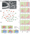
Figure 1
Figure 1. IIS network responds to UV-induced DNA damage during development and activation of DAF-16 that declines with aging
(A) Transcription-coupled repair defects lead to arrested somatic development. Worms were treated as L1 larvae and pictures taken 96h post treatment (wt, xpc-1, csb-1 with 60mJ/cm2; xpc-1;csb-1 with 10mJ/cm2). Note that while wt worms have developed into adult worms, xpc-1 mutants have completed somatic development but lack a germ line, csb-1 mutants are developmentally delayed while germ cells continued to divide while xpc-1;csb-1 double mutants have arrested both somatic and germ line development (representative pictures shown, experiment was repeated at least 3 times). (B) shows largest significantly overrepresented interaction network among differentially expressed genes upon UV treatment. Upregulated genes are depicted in red, downregulated genes in green. One link through non-transcriptionally regulated genes was permitted in the analysis. (C) DAF-16::GFP cellular localization upon UV treatment in larvae. L1 wt, xpc-1, xpa-1, csb-1 and csb-1;xpc-1 strains were irradiated with 120 mJ/cm2 and kept at 20°C (Average of n=3 independent experiments per strain and dose is shown, 10 individuals analysed per experiment; error bars=SEM). The graph shows the percentage of worms showing cytoplasmic, nuclear and partially nuclear DAF-16::GFP localization without treatment (control), and following UV irradiation. (D) DAF-16::GFP cellular localization upon UV treatment in adult worms. Wt and xpa-1 mutant are shown. Three replicates of 10 adult worms during the first, third, fifth, seventh and tenth day of adulthood were exposed to 150 mJ/cm2 and incubated at 20°C. In parallel, adult worms of the same age were heat shocked at 32°C for ten minutes and scored for the subcellular localization of the GFP signal (Average of n=3 independent experiments per strain and dose is shown, 10 individuals analysed per experiment; error bars=SEM).
Figure 2
Figure 2. Activation of DAF-16 promotes developmental growth upon UV-induced damage
daf-2 and age-1 mutants show reduced developmental arrest in a daf-16 dependent manner. L1 larvae were treated with UV and larval stages determined 48h post treatment. Animals that proceeded to L4 and young adulthood (YA) are scored together. Note that daf-16 single mutant alleles are analysed in suppl. Figure 2E. (Average of n=4 independent experiments per strain and dose is shown, >1450 individuals analysed per experiment; error bars=SD, *=p<0.05, **=p<0.01, ***=p<0.001, two-tailed T-Test, compared to wt).
Figure 3
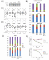
Figure 3. IIS mutants enhance somatic resistance to persistent DNA damage
(A) UV-induced DNA lesions remain unrepaired in xpa-1 and in xpa-1;daf-2 mutants. wt, xpa-1, and xpa-1;daf-2 were treated at L1 stage and CPD and 6-4PP lesions were measured by antibody staining in slot blots of worm extracts directly upon treatment with 40mJ/cm2 UV and 24h later (representative result is shown and the experiment was repeated 3 times) (B) L1 larvae were irradiated or mock treated and developmental stages evaluated 48h or 72h (lower panel) later (average of n=3 independent experiments per strain and dose is shown, >829 individuals analysed per experiment; error bars=SD; *=p<0.05, **=p<0.01, ***=p<0.001, two-tailed T-Test compared to wt) (C) Worms were isolated as L4s and irradiated 24hs later or mock treated and pharyngeal pumping rate per 10 seconds was determined on day 7 after the irradiation (representation in box-and-whisker graph with whiskers 1.5x IQR, ****=p<0.0001, two-tailed T-Test). (D) For body bends measurements, worms were isolated as L4s and irradiated 24hs later or mock treated and body bend rate per 10 seconds was determined on day 8 after the irradiation (representation in box-and-whisker graph with whiskers 1.5x IQR, ***=p<0.001, two-tailed T-Test). (E) L1 larvae were treated and developmental stages assessed 48h post treatment (average of n=3 independent experiments per strain and dose is shown, >3400 individuals analysed per experiment; error bars=SD, *=p<0.05, **=p<0.01, ***=p<0.001, two-tailed T-Test, compared to csb-1). (F) L4 larvae were irradiated and were allowed to lay eggs for 5 hours starting at 24h after treatment (F; upper panel; average of n=3 independent experiments per strain and dose, 3 individuals analysed per experiment, error bars=SD). Viable offspring was counted (F; lower panel) 48h post egg-laying (average of n=3 independent experiments per strain and dose shown, offspring of 3 parental individuals analysed per experiment; error bars=SD).
Figure 4
Figure 4. Distinct and similar responses to persistent DNA damage and starvation
Synchronized L1 worms were fed for 4 hours or left under starvation. Starvation responses were previously shown to be quickly reverted upon feeding. Fed wild type worms were treated with 60 mJ/cm2 UV and compared to non-UV treated controls. (A) xy blot of fold changes of significantly differentially expressed genes in UV treated and starved wt worms; linear regression is depicted in red. (B) Venn diagram showing overlap between significantly differentially expressed genes in response to starvation and UV treatment. (C) Functional clustering of genes that were up- or downregulated upon UV treatment (p<0.01, FC>±1.5) or starvation (p<0.01, FC>±2) reveals significantly overrepresented biological processes. (D) Synchronized L1 larvae were either fed for four hours or left under starvation and exposed to UV treatment. Upon UV treatment larvae were provided with food and developmental stages were assessed 48h post UV irradiation (average of n=3 independent experiments is shown, >796 individuals analysed per experiment; error bars=SD, ***=p<0.001, two-tailed T-Test).
Figure 5
Figure 5. The GATA transcription factor EGL-27 promotes developmental growth upon DNA damage
(A) Specifically 3 GATA motifs are significantly enriched in promoters of DAF-16 dependently induced genes in response to DNA damage. RSAT analysis identified three motifs that were significantly enriched in 1000 bp upstream sequences of 449 (RSAT eligible out of 514 total; see methods) genes that were significantly induced in daf-2 mutants (FC>1.5; p<0.01) upon UV and more strongly induced upon DNA damage in daf-2 vs. daf-2;daf-16, daf-2 vs. wt, and wt vs. daf-16. Motif comparisons with transcription factors databases identified significant match to GATA motifs. (B) egl-27(n170) and egl-27(ok1670) mutants are hypersensitive to larval arrest following DNA damage and rescued by transgenic egl-27::GFP expression. Synchronized L1 larvae were exposed to UV and larval stages assessed 48h post treatment (average of n=3 independent experiments per strain and dose is shown, >900 individuals analysed per experiment; error bars=SD *=p<0.05, **=p<0.01, ***=p<0.001, two-tailed T-Test).
Figure 6
Figure 6. The GATA transcription factor EGL-27 co-regulates DAF-16 target genes and acts downstream of DAF-2 to promote developmental growth in response to DNA damage
(A) egl-27 is required for DAF-16 target gene induction. Genes that were defined as DAF-16 dependently induced (see Figure legend 5A) were analysed by qPCR 6h post UV in wt, egl-27(n170), and egl-27(ok1670) mutants (average of n=3 independent experiments). (B) RNAi knockdown of “DNA damage effector downstream of DAF-16 and EGL-27” dde-1 and dde-2 by two independent RNAi constructs (RNAi #1, RNAi #2) and a genetic mutation in dde-3(gk696370) lead to enhanced UV sensitivity (dde-3 mutant was outcrossed prior to analysis; average of n=3 independent experiments per strain and dose is shown, >211 individuals analysed per experiment; error bars=SD; *=p<0.05, **=p<0.01, ***=p<0.001, two-tailed T-Test, compared to GFP RNAi or wt, respectively). (C) wt, daf-16, egl-27 and daf-16;egl-27 upon UV treatment were analysed 48h post UV (average of n=3 independent experiments per strain and dose is shown, >25 individuals analysed per experiment analysed; error bars=SD; *=p<0.05, **=p<0.01, ***=p<0.001, two-tailed T-Test compared to wt). (D) egl-27 suppresses UV tolerance of daf-2. Synchronized L1 larvae were UV treated and developmental stages assessed 48h post treatment (average of n=3 independent experiments per strain and dose is shown, >1738 individuals analysed per experiment; error bars=SD, *=p<0.05, **=p<0.01, ***=p<0.001, two-tailed T-Test compared to wt).
Figure 7
Figure 7. EGL-27 interacts with DAF-16 upon DNA damage
(A) DAF-16::GFP upon UV treatment co-localizes with EGL-27::GFP in the nucleus of intestinal cells. L1 synchronized larvae were fed and UV-treated with 150 mJ/cm2 and kept at 20°C, images were taken 6hs after. Upper panels show DAF-16::GFP worms, left panel non-UV treated and right panel a completely nuclear DAF-16::GFP UV-irradiated worm. Lower panels show EGL-27::GFP worms, left panel non-UV treated and right panel UV-irradiated (representative images shown; experiment repeated at least 3 times). (B) EGL-27 interacts with DAF-16 (left panel). HEK293T cells were transiently transfected and after immunoprecipitation (IP) with FLAG (M2) beads, Western blot analysis revealed that DAF-16.V5 coprecipitated with EGL-27.FLAG but not with a control protein (EPS) (representative result of 4 independent experiments). Interaction with DAF-16 is mediated by the C-terminal part of EGL-27 (right panel). HEK293T cells were transiently transfected and after IP with FLAG (M2) beads, Western blot analysis revealed that DAF-16.V5 co-precipitated with the EGL-27 C-terminus but not with an N-terminal truncation or a control protein (EPS) (representative result of 4 independent experiments). (C) Model for the differential consequences of DAF-16 activation upon persistent DNA damage and upon starvation. In response to persistent DNA lesions in somatic tissues DAF-16 activation results in induction of somatic growth genes, which are repressed by DAF-16 upon starvation, through recognition of DAE promoter elements through the GATA transcription factor EGL-27. In both cases stress response genes are induced through recognition of DBE motifs leading to enhanced tissue maintenance.

References

    1. Loeb LA, Harris CC. Advances in chemical carcinogenesis: a historical review and prospective. Cancer Res. 2008;68:6863–6872. - PMC - PubMed
    1. Schumacher B, Garinis GA, Hoeijmakers JHJ. Age to survive: DNA damage and aging. Trends Genet. 2008;24:77–85. - PubMed
    1. Cleaver JE, Lam ET, Revet I. Disorders of nucleotide excision repair: the genetic and molecular basis of heterogeneity. Nat Rev Genet. 2009;10:756–768. - PubMed
    1. Wolters S, Schumacher B. Genome maintenance and transcription integrity in aging and disease. Front Genet. 2013;4:19. - PMC - PubMed
    1. Bartek J, Lukas J. DNA damage checkpoints: from initiation to recovery or adaptation. Curr. Opin. Cell Biol. 2007;19:238–245. - PubMed

Additional References

    1. Brenner S. The genetics of Caenorhabditis elegans. Genetics. 1974;77:71–94. - PMC - PubMed
    1. Pfaffl MW. A new mathematical model for relative quantification in real-time RT-PCR. Nucleic Acids Res. 2001;29:e45. - PMC - PubMed
    1. Furuyama T, Nakazawa T, Nakano I, Mori N. Identification of the differential distribution patterns of mRNAs and consensus binding sequences for mouse DAF-16 homologues. Biochem. J. 2000;349:629–634. - PMC - PubMed
    1. Sakurai H, Enoki Y. Novel aspects of heat shock factors: DNA recognition, chromatin modulation and gene expression. FEBS J. 2010;277:4140–4149. - PubMed
    1. Sinclair J, Hamza I. A novel heme-responsive element mediates transcriptional regulation in Caenorhabditis elegans. J Biol Chem. 2010;285:39536–39543. - PMC - PubMed

Publication types

MeSH terms