. 2014 Dec;16(12):1146-56.
doi: 10.1038/ncb3070. Epub 2014 Nov 24.

SOX17 links gut endoderm morphogenesis and germ layer segregation

Affiliations

SOX17 links gut endoderm morphogenesis and germ layer segregation

Manuel Viotti et al. Nat Cell Biol. 2014 Dec.

Abstract

Gastrulation leads to three germ layers--ectoderm, mesoderm and endoderm--that are separated by two basement membranes. In the mouse embryo, the emergent gut endoderm results from the widespread intercalation of cells of two distinct origins: pluripotent epiblast-derived definitive endoderm (DE) and extra-embryonic visceral endoderm (VE). Here we image the trajectory of prospective DE cells before intercalating into the VE epithelium. We show that the transcription factor SOX17, which is activated in prospective DE cells before intercalation, is necessary for gut endoderm morphogenesis and the assembly of the basement membrane that separates gut endoderm from mesoderm. Our results mechanistically link gut endoderm morphogenesis and germ layer segregation, two central and conserved features of gastrulation.


Figures

Figure 1
Figure 1. DE cells originate in the posterior epiblast and migrate with the wings of mesoderm before egressing into the emVE epithelium
(a) Schematic depicting the electroporation and time-lapse imaging procedure. (b–e) Interior rendered views from a time-lapse. (b′–e′) Surface rendered views from a time-lapse (b–e). (f–i) Afp::GFP VE-reporter embryos showing progression of emVE dispersal from pre-dispersal (PS stage, E6.25) to late/completed dispersal (LB/EHF stage, E7.5) stage. (f′–i′) Transverse sections through Afp::GFP embryos in (f–i). (j and j′) Whole mount view and transverse section of Fgf8 mutant, transgenic for the Afp::GFP VE-reporter, showing accumulation of cells in the area of the primitive streak and no emVE dispersal. ps, primitive streak; emVE, embryonic visceral endoderm; epi, epiblast; exVE, extraembryonic visceral endoderm; mes, mesoderm; A, anterior; D, distal; L, left; P, posterior; Pr, proximal; R, right; PS, pre-streak; LS, late streak; OB, no bud; LB, late bud; EHF, early head-fold. Scale bars = 100 μm. See also Supplementary Fig. 1 and Supplementary Videos 1–5.
Figure 2
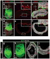
Figure 2. SOX17 marks DE cells prior to, during, and after egression and is required for egression event
(a) Whole mount image of immuno-fluorescence for SOX17 in an Afp::GFP VE-reporter embryo at the early emVE dispersal stage. (b and c) Sections through embryo in (a) indicate two different expression levels of SOX17 during early emVE dispersal. EmVE cells display low levels of SOX17 (pink arrowheads), and DE cells in the process of egressing display high levels of SOX17 (orange arrowheads). Orange asterisks mark the leading edge of the mesoderm. (d) Whole mount image of immuno-fluorescence for SOX17 in an Afp::GFP VE-reporter embryo at the late/completed emVE dispersal stage. (e and f) Sections through embryo in (d) indicate equal levels of SOX17 expression in all cells of the gut endoderm layer, regardless if emVE-derived (GFP-positive) or of the DE lineage (GFP-negative). (g) Sox17 mutant Afp::GFP VE-reporter embryo at the LS stage displays a solid layer of GFP-positive cells on its distal surface. (h) Sox17 mutant Afp::GFP VE-reporter embryo at the LB/EHF stage showing a uniform layer of GFP-positive cells on its distal surface, suggesting continuous failure of DE-cell egression. Limited egression of DE cells into the emVE has occurred anteriorly in the area around the prospective foregut invagination (white arrowhead). (i and j) Low and high magnifications of sections through Sox17 mutant embryo in (h) show wings of mesoderm that have completed anterior migration and a solid GFP-positive layer on the surface of the embryo, representing the undispersed emVE epithelium. ps, primitive streak; end, endoderm; epi, epiblast; mes, mesoderm; A, anterior; D, distal; L, left; P, posterior; Pr, proximal; R, right; LS, late streak; LB, late bud; EHF, early head-fold. Scale bars = 100 μm. See also Supplementary Fig. 2.
Figure 3
Figure 3. FOXA2 is not required for DE cell egression or SOX17 expression
(a–d) Whole mount images of Foxa2 mutant expressing the Afp::GFP VE-reporter at the LB/EHF (E7.5) stage, with immuno-fluorescence for SOX17. Note that the midline has failed to undergo correct formation (orange arrowhead), but the emVE has undergone dispersal (white arrowhead). A, anterior; D, distal; P, posterior; Pr, proximal; LB, late bud; EHF, early head-fold. Scale bar = 100 μm.
Figure 4
Figure 4. Cells failing to egress remain within the mesodermal wings
(a–d) Low and high magnifications of sections through a LB/EHF stage (E7.5) Sox17GFP/+ embryo with immuno-fluorescence for the endoderm marker FOXA2, indicating a single layer of GFP-positive cells on the surface of the embryo (all cells of the gut endoderm), uniformly expressing FOXA2. (a′–d′) A Sox17GFP/GFP embryo additionally displays some GFP-positive cells embedded within the mesodermal layer and do not express FOXA2(orange arrowheads). (e–h) Low and high magnification views of sections through a Sox17GFP/+ embryo depicting immuno-fluorescence for the epithelial marker E-CADHERIN (E-CAD). Fluorescent signal is present between cells in the epiblast layer and in the gut endoderm, which is GFP-positive. (e′–h′) A Sox17GFP/GFP embryo additionally displays some GFP-positive cells within the wings of mesoderm. These ‘non-egressed’ cells show low levels of cytoplasmic E-CAD (orange arrowheads). (i–l) Low and high magnifications of sections through a Sox17GFP/+ embryo depicting immuno-fluorescence for the mesenchymal marker N-CADHERIN (N-CAD). N-CAD stain is present between cells of the wings of mesoderm, and absent from cells of the epiblast or the gut endoderm, which is GFP-positive. (i′–l′) A Sox17GFP/GFP embryo additionally displays some GFP-positive cells within the wings of mesoderm. These ‘non-egressed’ cells show N-CAD at their interface with neighbouring mesodermal cells (orange arrowheads). ps, primitive streak; emVE, embryonic visceral endoderm; end, endoderm; epi, epiblast; exVE, extraembryonic visceral endoderm; mes, mesoderm; A, anterior; D, distal; L, left; P, posterior; Pr, proximal; R, right; LB, late bud; EHF, early head-fold. Scale bars = 100 μm. See also Supplementary Fig. 3.
Figure 5
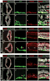
Figure 5. Formation of basement membrane at mesoderm-endoderm interface does not occur in Sox17 mutants
(a–l′) Immuno-fluorescence for the BM protein FN-1 in Afp::GFP VE-reporter embryos. (a–c′) At the ES stage (E6.75), shortly after the wings of mesoderm have begun their anterior migration (leading tips indicated by orange asterisks), a single continuous signal for FN-1 is present between epiblast and the GFP-positive emVE. (d–f′) During early emVE dispersal, one BM is visible anterior to the leading tips of the wings of mesoderm (orange asterisks), and two BMs in regions where the wings of mesoderm are present. The BM between mesoderm and endoderm is heavily fenestrated. (g–i′) FN-1 at mid-emVE dispersal identifies two BMs where the wings of mesoderm are present. (j–l′) FN-1 at the late/completed emVE dispersal stage identifies two solid BMs separating germ layers. (m–p) Double stains in Afp::GFP VE-reporter embryo shows egressing DE cells with high levels of SOX17 in the process of inserting between emVE cells displaying FN-1 basally. EmVE cells never display FN-1 at their interface with egressing DE cells. (q–t) Stained Afp::GFP VE-reporter wild-type embryo showing egressing DE cells (orange asterisks) always display FN-1 basally. (q′–t′) In the Sox17 mutant, the interface between mesoderm and emVE only displays faint punctate FN-1 fluorescent signal. (u and u′) Digital quantitation of FN-1 fluorescent signal indicating two peaks in the wild-type and only one peak in the Sox17 mutant. (v–y) Stained Afp::GFP VE-reporter wild-type embryo showing egressing DE cells (orange asterisks) always display LAMA-1 basally. (v′–y′) In Sox17 mutants, the interface between mesoderm and emVE displays punctate LAMA-1 fluorescent signal, similarly to the puncta interspersed between cells of the wings of mesoderm of the wild-type. (z and z′) Digital quantitation of fluorescent signal indicating two peaks for LAMA-1 in the wild-type, and only one in Sox17 mutant. ps, primitive streak; emVE, embryonic visceral endoderm; end, endoderm; epi, epiblast; mes, mesoderm; A, anterior; D, distal; L, left; P, posterior; Pr, proximal; R, right; PS, pre-streak; LS, late streak; OB, no bud; LB, late bud; EHF, early head-fold. Scale bars = 20 μm in panel m, 100 μm in all other panels. See also Supplementary Fig. 4 and Supplementary Video 6.
Figure 6
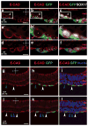
Figure 6. E-CADHERIN distribution dynamics in DE and emVE cells
(a–f) High magnifications of sectioned OB (E7.25) Afp::GFP VE-reporter wild-type embryos stained for E-CAD and SOX17. Strong white signal indicates egressing DE-cells (orange asterisks) in the process of inserting between GFP-positive emVE cells (pink asterisks). Note the polarization of E-CAD signal in egressing cells, with strong localization (pink arrowheads) on their cell membrane interfacing with emVE cells. (g–l) E-CAD stain in EHF (E7.75) and LHF (E8.0) stage Afp::GFP VE-reporter wild-type embryos. Note the strong localization of E-CAD in the gut endoderm between fully egressed DE cells (blue arrowheads) as well as at the interface between DE and GFP-positive emVE-derived cells (white arrowheads). end, endoderm; epi, epiblast; mes, mesoderm; A, anterior; L, left; P, posterior; R, right; OB, no bud; EHF, early head-fold; LHF, late head-fold. Scale bars = 20μm. See also Supplementary Fig. 5.
Figure 7
Figure 7. DE cells in Sox17 mutants fail to epithelialize
(m–o) Digital quantitation of fluorescent intensities. In six or more wild-type embryos, we measured 50 egressing cells and 50 inner cells (deeper in the mesoderm layer). In >6 Sox17 mutants, we measured 25 cells at the interface with the VE and 25 inner cells. Dot plot of two measurements made for each cell; one for the cell’s apex facing the embryo surface (blue triangles) and the other for the apex facing the embryo cavity (purple dots) (see Supplementary Fig. 7). Numbers above the graph indicate the instances in which the measurement on the side of the embryo surface was higher or lower than the measurement on the side of the embryo cavity. Histogram indicates the mean ratio between higher and lower value for each cell type (n=50 for wild-type cells, n=25 in mutant cells). Error bars represent standard deviation. ps, primitive streak; end, endoderm; epi, epiblast; mes, mesoderm; A, anterior; L, left; P, posterior; R, right; OB, no bud; s, side of embryo surface; c, side of embryo cavity. Scale bars = 100 μm. See also Supplementary Figs. 6 and 7.
Figure 8
Figure 8. Working model of cell behaviors during gut endoderm morphogenesis and germ layer segregation in mice
The gut endoderm forms by widespread intercalation between embryonic and extra-embryonic endoderm, which occurs concomitantly with the assembly of a BM at its interface with the mesoderm. Gastrulation transforms a two-layered to a three-tissue layered tissue configuration. Epiblast cells ingress and undergo EMT at the primitive streak. They emerge as mesoderm or gut endoderm. SOX17 orchestrates a mesenchymal-to-epithelial transition (MET) of DE cells at the interface with the VE, in which DE cells become polarized, enrich ECM receptors, and assemble BM components basally. The overlying VE layer transiently moderates its epithelial characteristics, facilitating DE cell egression. The emergent gut endoderm (composed of VE and DE cells) subsequently reinforces the underlying BM, segregating mesoderm from gut endoderm tissue layers.

Comment in

References

    1. Nowotschin S, Hadjantonakis AK. Cellular dynamics in the early mouse embryo: from axis formation to gastrulation. Curr Opin Genet Dev. 2010;20:420–427. - PMC - PubMed
    1. Arnold SJ, Robertson EJ. Making a commitment: cell lineage allocation and axis patterning in the early mouse embryo. Nat Rev Mol Cell Biol. 2009;10:91–103. - PubMed
    1. Rivera-Perez JA, Hadjantonakis AK. The Dynamics of Morphogenesis in the Early Mouse Embryo. Cold Spring Harbor perspectives in biology. 2014;a015867 doi: 10.1101/cshperspect.a015867. - DOI - PMC - PubMed
    1. Zorn AM, Wells JM. Vertebrate endoderm development and organ formation. Annu Rev Cell Dev Biol. 2009;25:221–251. - PMC - PubMed
    1. Viotti M, Nowotschin S, Hadjantonakis AK. Afp::mCherry, a red fluorescent transgenic reporter of the mouse visceral endoderm. Genesis. 2011;49:124–133. - PMC - PubMed

Publication types

MeSH terms