. 2014 Nov 24;9(11):e111911.
doi: 10.1371/journal.pone.0111911. eCollection 2014.

Elevated expression of AKR1C3 increases resistance of cancer cells to ionizing radiation via modulation of oxidative stress

Affiliations

Elevated expression of AKR1C3 increases resistance of cancer cells to ionizing radiation via modulation of oxidative stress

Wei Xiong et al. PLoS One. .

Abstract

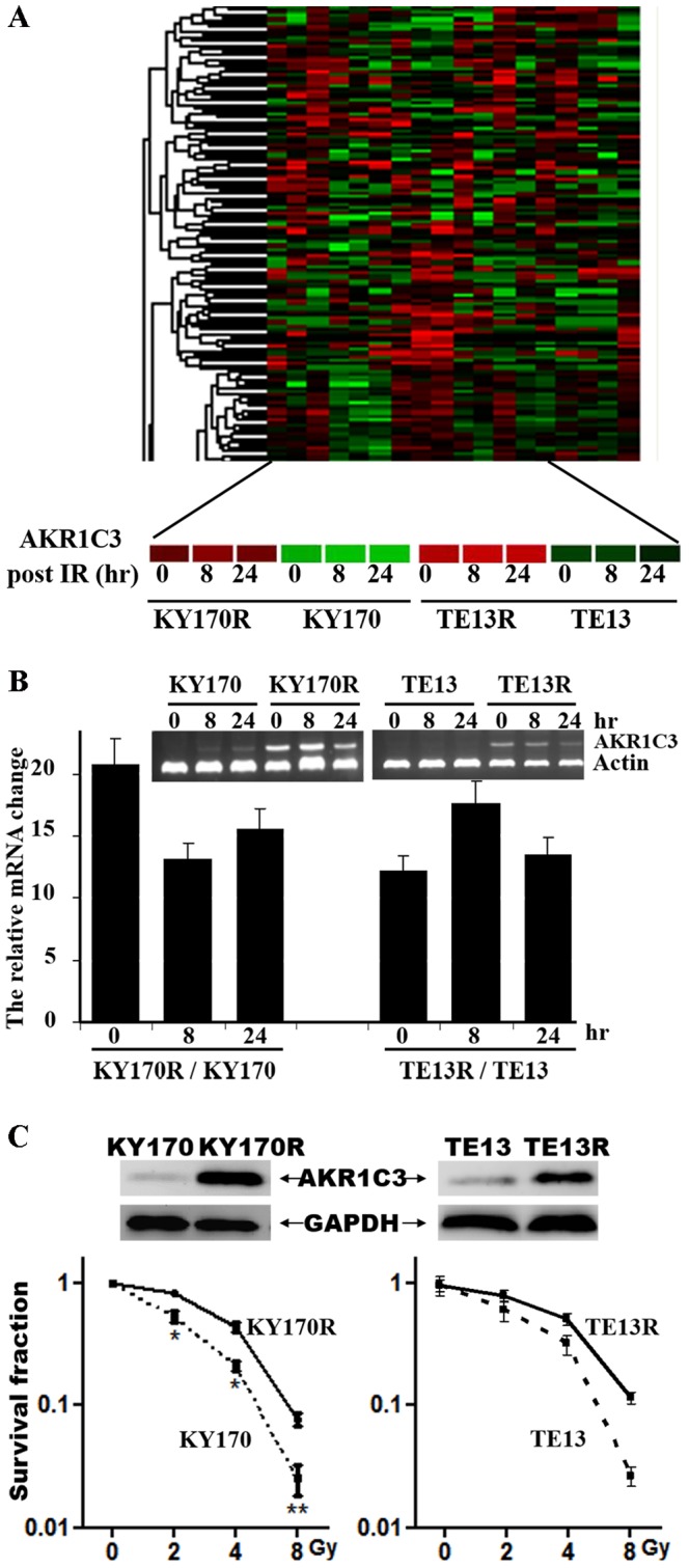
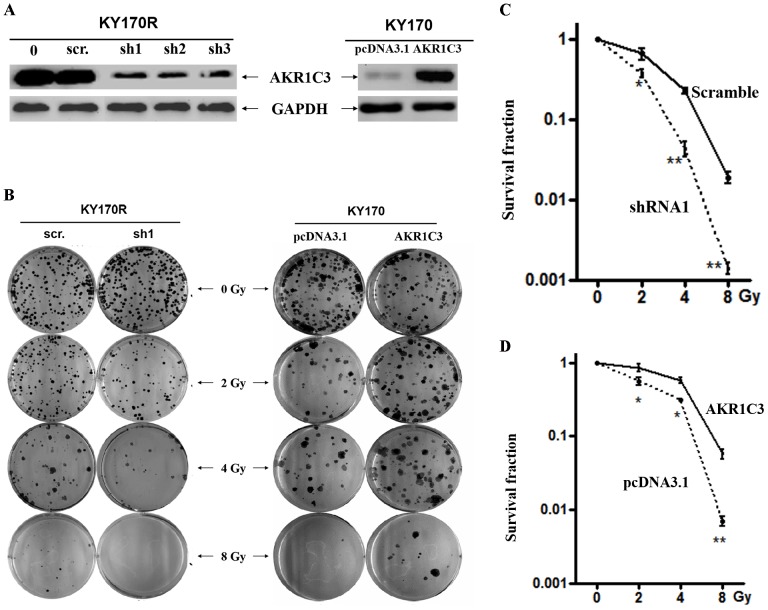
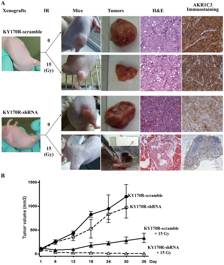
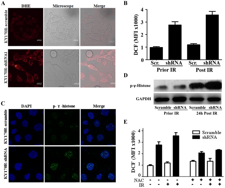
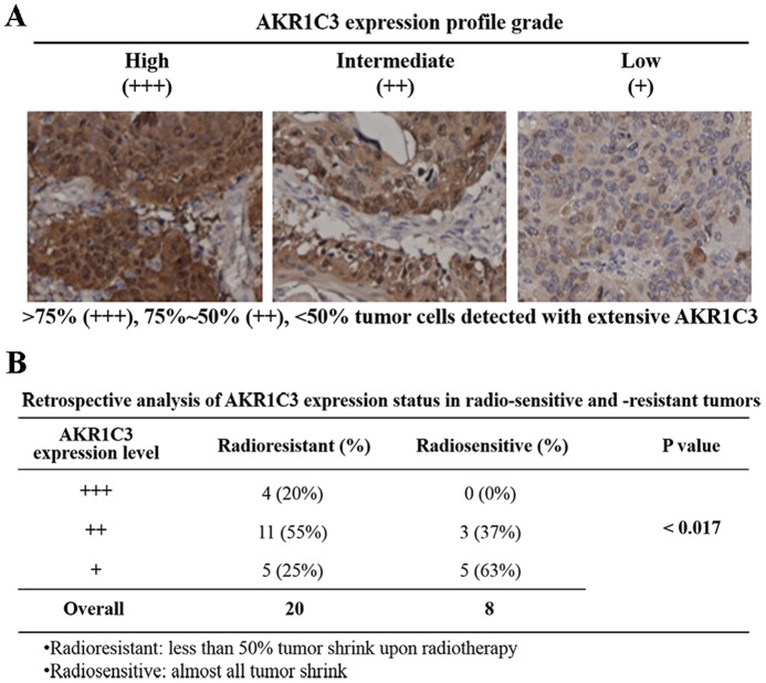
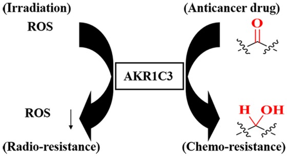
With the aim to elucidate the etiology of radioresistance, we explored the genetic alterations in non-radioresistant vs. resistant esophageal cancer cells acquired by long-term fractionated radiation. We found AKR1C3, an aldo-keto reductase expressed seldom in most human tissues, expressed higher in radioresistance-acquired cells. Suppression of AKR1C3 via RNAi or its chemical inhibitors restored the sensitivity of the acquired tumor cells and xenograft BALB/c nude mice to ionizing radiation (IR). Cellular monitoring of the oxidative stress in the AKR1C3-elevated cells indicated that IR-induced ROS accumulation and the concomitant DNA damage was significantly alleviated, and such protective consequence disappeared upon AKR1C3 knockdown. These findings uncover the potential involvement of AKR1C3 in removal of cellular ROS and explain, at least partially, the acquired radioresistance by AKR1C3 overexpression. A retrospective analysis of esophageal carcinomas also indicated a significant expression of AKR1C3 in radio-resistant but not radio-sensitive surgical samples. Our study may provide a potential biomarker for predicting prognosis of radiotherapy and even direct a targeted therapy for esophageal cancer and other tumors.


Conflict of interest statement

Competing Interests: The authors have declared that no competing interests exist.

Figures

Figure 1
Figure 1. Profiling of AKR1C3 in radio-resistant and -sensitive cancer cells.
(A) Genome-wide profiling of gene expression in radioresistant KY170R and TE13R versus radiosensitive KY170 and TE13, respectively, at 0, 8 h and 24 h after irradiation. (B) Validation of the elevated expression of AKR1C3 at the mRNA level in KY170R vs. KY170 and TE13R vs. TE13 cells prior to and 8 and 24 hr after irradiation as determined by RT-PCR. (C) Validation of elevated expression of AKR1C3 in radioresistant KY170R determined by Western blot and the sensitivity of KY170 and KY170R cells to irradiation as determined by a colony-formation assay.
Figure 2
Figure 2. Cell-based evaluation of the role of AKR1C3 on the response of esophageal cancer cells to irradiation.
(A) Representative observations of stable knockdown of AKR1C3 in KY170R cells by three independent shRNAs and overexpression of AKR1C3 in KY170 cells. (B) The effects of AKR1C3 knockdown in KY170R cells and AKR1C3 overexpression in KY170 on the outcome of irradiation, dosed from 0 to 8 Gy, determined by colony-formation assay. A scramble shRNA (scr.) and the empty pcDNA3.1 vector acted as a negative control, respectively. (C) The survival curves for comparisons of the effect of AKR1C3 on the sensitivity of KY170R cells (knockdown) and (D) KY170 cells (overexpression) to irradiation, as determined by colony-formation assays.
Figure 3
Figure 3. Xenograft-based evaluation of AKR1C3 elevation on the response of esophageal cancers to irradiation.
(A) Representative observations of the growth of xenograft KY170R-scramble and KY170R-shRNA1 tumors, and the responses of these tumors to a single dose (15 Gy) of irradiation in terms of tumor volume, HE staining and immunochemical staining of AKR1C3. (B) The time course of growth of KY170R-scramble and KY170R-shRNA1 xenograft tumors with or without a local IR treatment.
Figure 4
Figure 4. Mechanistic exploration of AKR1C3 as a cellular factor for protecting cells from irradiation damage.
(A) Representative microscopic view of the accumulation of ROS in KY170R-scramble v. KY170R-shRNA1 cells stained by DHE. (B) Quantifications of the ROS levels in KY170R-scramble v. KY170R-shRNA1 cells prior to and 24 h after IR at a dose of 4 Gy. (C) Representative microscopic view of phospho-γ-histone foci in KY170R-scramble v. KY170R-shRNA1 cells. (D) Characterizations of phospho-γ-histone in tested cells by Western blotting. (E) N-Acetyl cysteine (NAC)-mediated scavenging of ROS at a IR dose of 4 Gy. Each quantitative measurement was carried out in triplicate and repeated at least twice with an error bar less than 25%.
Figure 5
Figure 5. Retrospective analysis of the relevance of AKR1C3 expression and the outcome of IR in esophageal carcinomas.
(A) Subjective levels of expression of AKR1C3, namely, high (+++), intermediate (++), low or none (+/−), in esophageal carcinomas. (B) Levels of expression of AKR1C3 in esophageal carcinoma from 28 patients who were congenitally sensitive or resistant to IR.
Figure 6
Figure 6. A proposed mechanism of AKR1C3-mediated radioresistance and certain cases of chemo-resistance via its reductive capability.

References

    1. Bernier J, Hall EJ, Giaccia A (2004) Radiation oncology: a century of achievements. Nat Rev Cancer 4: 737–747. - PubMed
    1. Riley PA (1994) Free radicals in biology: oxidative stress and the effects of ionizing radiation. Int J Radiat Biol 65: 27–33. - PubMed
    1. Majima HJ, Indo HP, Tomita K, Suenaga S, Motoori S, et al.. (2006). Intracellular oxidative stress caused by ionizing radiation. In: Singh KK (ed.) Oxidative Stress, Disease and Cancer. London: Imperial College Press. pp.61–83.
    1. Spitz DR, Azzam EI, Li JJ, Gius D (2004) Metabolic oxidation/reduction reactions and cellular responses to ionizing radiation: A unifying concept in stress response biology. Cancer And Metastasis Reviews 23: 311–322. - PubMed
    1. Roos WP, Kaina B (2006) DNA damage-induced cell death by apoptosis. Trends In Molecular Medicine 12: 440–450. - PubMed

Publication types

MeSH terms

Substances

Associated data