

Resibufogenin and cinobufagin activate central neurons through an ouabain-like action
- PMID: 25420080
- PMCID: PMC4242513
- DOI: 10.1371/journal.pone.0113272
Resibufogenin and cinobufagin activate central neurons through an ouabain-like action
Abstract
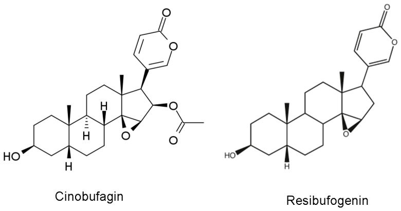
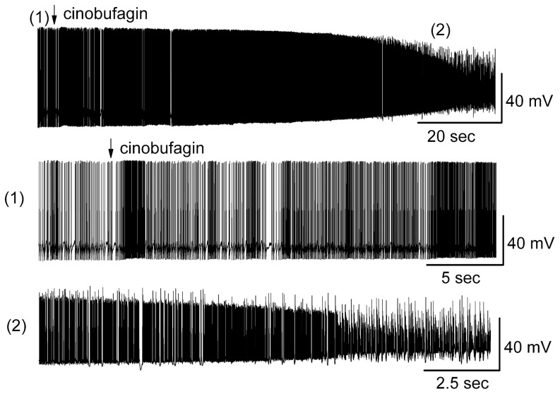
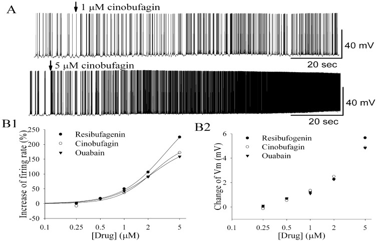
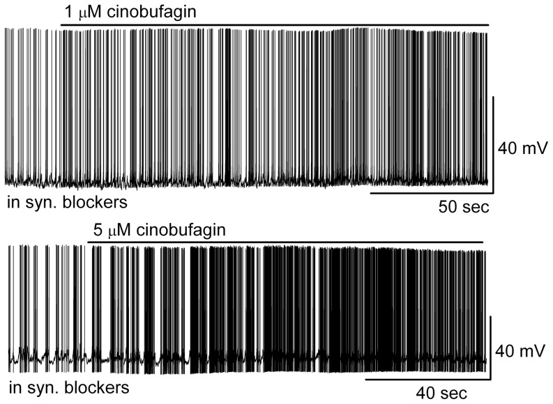
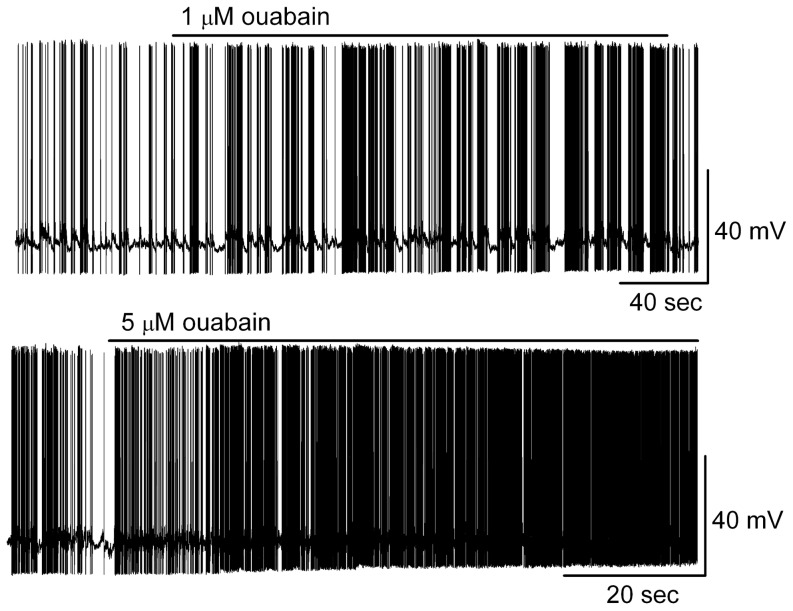
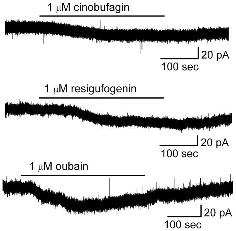
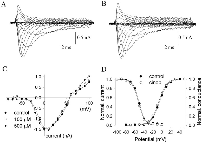
Cinobufagin and resibufogenin are two major effective bufadienolides of Chan su (toad venom), which is a Chinese medicine obtained from the skin venom gland of toads and is used as a cardiotonic and central nervous system (CNS) respiratory agent, an analgesic and anesthetic, and as a remedy for ulcers. Many clinical cases showed that Chan su has severe side-effects on the CNS, causing shortness of breath, breathlessness, seizure, coma and cardiac arrhythmia. We used whole-cell recordings from brain slices to determine the effects of bufadienolides on excitability of a principal neuron in main olfactory bulb (MOB), mitral cells (MCs), and the cellular mechanism underlying the excitation. At higher concentrations, cinobufagin and resibufogenin induced irreversible over-excitation of MCs indicating a toxic effect. At lower concentrations, they concentration-dependently increased spontaneous firing rate, depolarized the membrane potential of MCs, and elicited inward currents. The excitatory effects were due to a direct action on MCs rather than an indirect phasic action. Bufadienolides and ouabain had similar effects on firing of MCs which suggested that bufadienolides activated neuron through a ouabain-like effect, most likely by inhibiting Na+/K+-ATPase. The direct action of bufadienolide on brain Na+ channels was tested by recordings from stably Nav1.2-transfected cells. Bufadienolides failed to make significant changes of the main properties of Nav1.2 channels in current amplitude, current-voltage (I-V) relationships, activation and inactivation. Our results suggest that inhibition of Na+/K+-ATPase may be involved in both the pharmacological and toxic effects of bufadienolide-evoked CNS excitation.
Conflict of interest statement
Figures
Similar articles
-
Effects of Resibufogenin and Cinobufagin on voltage-gated potassium channels in primary cultures of rat hippocampal neurons.Toxicol In Vitro. 2011 Dec;25(8):1644-53. doi: 10.1016/j.tiv.2011.07.001. Epub 2011 Jul 21. Toxicol In Vitro. 2011. PMID: 21798339
-
Effects of resibufogenin on voltage-gated sodium channels in cultured rat hippocampal neurons.Neurosci Lett. 2011 Aug 26;501(2):112-6. doi: 10.1016/j.neulet.2011.06.059. Epub 2011 Jul 8. Neurosci Lett. 2011. PMID: 21763753
-
Bufadienolides from amphibians: A promising source of anticancer prototypes for radical innovation, apoptosis triggering and Na+/K+-ATPase inhibition.Toxicon. 2017 Mar 1;127:63-76. doi: 10.1016/j.toxicon.2017.01.004. Epub 2017 Jan 7. Toxicon. 2017. PMID: 28069354 Review.
-
Conformational states of the pig kidney Na+/K+-ATPase differently affect bufadienolides and cardenolides: A directed structure-activity and structure-kinetics study.Biochem Pharmacol. 2020 Jan;171:113679. doi: 10.1016/j.bcp.2019.113679. Epub 2019 Oct 24. Biochem Pharmacol. 2020. PMID: 31669257
-
Toad venom bufadienolides and bufotoxins: An updated review.Drug Dev Res. 2023 Aug;84(5):815-838. doi: 10.1002/ddr.22072. Epub 2023 May 8. Drug Dev Res. 2023. PMID: 37154099 Review.
Cited by
-
Cinobufagin: Unveiling the hidden bufadienolide's promise in combating alimentary canal cancer development and progression - a comprehensive review.Naunyn Schmiedebergs Arch Pharmacol. 2025 Jul;398(7):8075-8089. doi: 10.1007/s00210-025-03902-9. Epub 2025 Feb 20. Naunyn Schmiedebergs Arch Pharmacol. 2025. PMID: 39976716 Review.
-
Resibufogenin: An Emerging Therapeutic Compound with Multifaceted Pharmacological Effects - A Comprehensive Review.Med Sci Monit. 2024 Feb 19;30:e942783. doi: 10.12659/MSM.942783. Med Sci Monit. 2024. PMID: 38369741 Free PMC article. Review.
-
Resibufogenin Targets the ATP1A1 Signaling Cascade to Induce G2/M Phase Arrest and Inhibit Invasion in Glioma.Front Pharmacol. 2022 May 17;13:855626. doi: 10.3389/fphar.2022.855626. eCollection 2022. Front Pharmacol. 2022. PMID: 35656311 Free PMC article.
-
The spinal microglial IL-10/β-endorphin pathway accounts for cinobufagin-induced mechanical antiallodynia in bone cancer pain following activation of α7-nicotinic acetylcholine receptors.J Neuroinflammation. 2020 Feb 29;17(1):75. doi: 10.1186/s12974-019-1616-z. J Neuroinflammation. 2020. PMID: 32113469 Free PMC article.
-
Aitongxiao improves pain symptoms of rats with cancer pain by reducing IL-1, TNF-α, and PGE2.Int J Clin Exp Pathol. 2021 Jan 1;14(1):133-139. eCollection 2021. Int J Clin Exp Pathol. 2021. PMID: 33532031 Free PMC article.
References
-
- Hong Z, Chan K, Yeung HW (1992) Simultaneous determination of bufadienolides in traditional Chinese medicine preparations, Liu-Shen-Wan by liquid chromatography. J Pharm Pharmacol 44:1023–1026. - PubMed
-
- Song H, Guo T, Bi K, Wang H, Zhang R (2000) Determination of resibufogenin and cinobufagin in heart-protecting musk pill by HPLC. Biomed Chromatogr 14:130–132. - PubMed
-
- Danchuk S, Sukhanov S, Horvat D, Uddin MN, Puschett JB (2008) Effects of resibufogenin in experimental hypertension. Am J Nephrol 28:8–13. - PubMed
-
- Dasgupta A, Biddle D, Wells A, Datta P (2000) Positive and negative interference of Chinese medicine Chan Su in serum digoxin measurement: elimination of interference by using a monoclonal chemiluminescent digoxin assay or monitoring free digoxin concentration. Am J Clin Pathol 114:174–179. - PubMed
Publication types
MeSH terms
Substances
Grants and funding
LinkOut - more resources
Full Text Sources
Other Literature Sources
