. 2014 Nov 24;9(11):e113423.
doi: 10.1371/journal.pone.0113423. eCollection 2014.

gone early, a novel germline factor, ensures the proper size of the stem cell precursor pool in the Drosophila ovary

Affiliations

gone early, a novel germline factor, ensures the proper size of the stem cell precursor pool in the Drosophila ovary

Shinya Matsuoka et al. PLoS One. .

Abstract

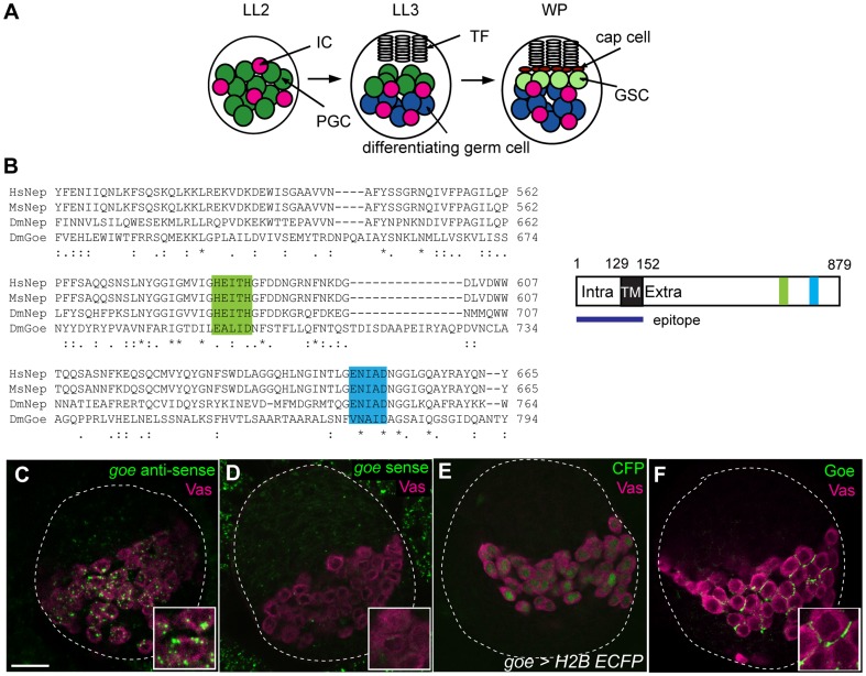
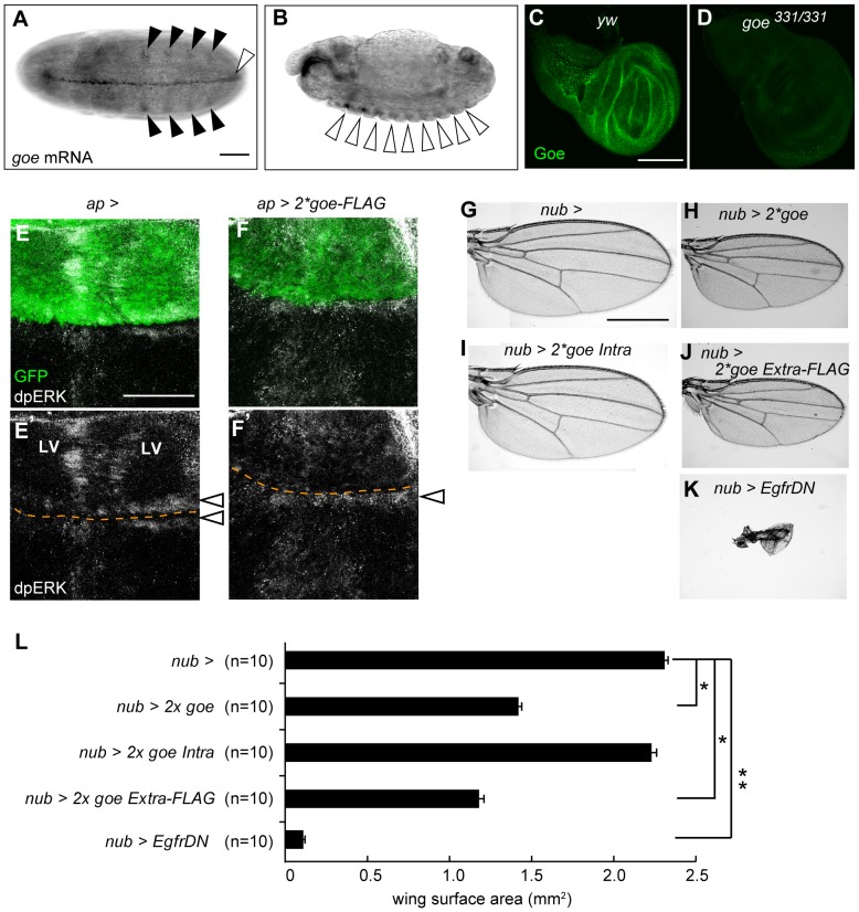
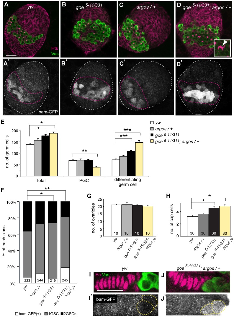
In order to sustain lifelong production of gametes, many animals have evolved a stem cell-based gametogenic program. In the Drosophila ovary, germline stem cells (GSCs) arise from a pool of primordial germ cells (PGCs) that remain undifferentiated even after gametogenesis has initiated. The decision of PGCs to differentiate or remain undifferentiated is regulated by somatic stromal cells: specifically, epidermal growth factor receptor (EGFR) signaling activated in the stromal cells determines the fraction of germ cells that remain undifferentiated by shaping a Decapentaplegic (Dpp) gradient that represses PGC differentiation. However, little is known about the contribution of germ cells to this process. Here we show that a novel germline factor, Gone early (Goe), limits the fraction of PGCs that initiate gametogenesis. goe encodes a non-peptidase homologue of the Neprilysin family metalloendopeptidases. At the onset of gametogenesis, Goe was localized on the germ cell membrane in the ovary, suggesting that it functions in a peptidase-independent manner in cell-cell communication at the cell surface. Overexpression of Goe in the germline decreased the number of PGCs that enter the gametogenic pathway, thereby increasing the proportion of undifferentiated PGCs. Inversely, depletion of Goe increased the number of PGCs initiating differentiation. Excess PGC differentiation in the goe mutant was augmented by halving the dose of argos, a somatically expressed inhibitor of EGFR signaling. This increase in PGC differentiation resulted in a massive decrease in the number of undifferentiated PGCs, and ultimately led to insufficient formation of GSCs. Thus, acting cooperatively with a somatic regulator of EGFR signaling, the germline factor goe plays a critical role in securing the proper size of the GSC precursor pool. Because goe can suppress EGFR signaling activity and is expressed in EGF-producing cells in various tissues, goe may function by attenuating EGFR signaling, and thereby affecting the stromal environment.


Conflict of interest statement

Competing Interests: The authors have declared that no competing interests exist.

Figures

Figure 1
Figure 1. Gone early, a non-peptidase homologue of Neprilysin metalloendopeptidases, is expressed in germline cells of LL3 ovaries.
(A) Key stages of PGC development. LL2, LL3, and WP are late second instar larval stage (72 h AEL [after egg laying]), late third instar larval stage (114 h AEL), and white pupal stage (120 h AEL), respectively. (B) Alignment of zinc-binding motifs of Gone early (Goe) with those of Neprilysin (Nep) from Homo sapiens (Hs), Mus musculus (Ms), and Drosophila melanogaster (Dm). The alignment was generated using the ClustalW algorithm. The symbols under the residues show Gonnet PAM 250 matrix scores: *, perfect match;: (colon),>0.5;. (period), ≤0.5. Amino-acid similarities between Goe and Hs, Ms, and Dm Nep are 38%, 37%, and 39%, respectively (between Hs Nep and Dm Nep: 59%). Two motifs are critical for the enzymatic activity of Neprilysin : HExxH (green), which contains two His residues that serve as zinc ligands and a Glu residue functioning in catalysis; and ExxA/GD (blue), which contains a Glu that serves as the third zinc ligand. Gone early lacks both of these motifs. (C–F) All confocal images depict LL3 ovaries. (C) An ovary stained for gone early (goe) mRNA and the germline marker Vasa (Vas, magenta). goe mRNA was detected in the germline. (D) Sense probe control. Insets in C and D show magnified views of germ cells. (E) An ovary stained for CFP (green, goe>H2B-ECFP) and Vasa (magenta). CFP expression driven by goe-gal4 (goe>H2B-ECFP) was found in the germline alone. For the enhancer element of goe-gal4, see Figure 3A. (F) An ovary stained with antibody against the intracellular and transmembrane domains of Goe (green) and anti-Vasa antibody (magenta). Anti-Vasa stained the cytoplasm of germ cells, whereas anti-Goe stained outside of germ cell cytoplasm, indicating that Goe was localized to the plasma membrane of germ cells. Goe was not distributed evenly on the germ cell plasma membrane, but was localized to sub-compartments of the membrane where neighboring germ cells face each other. The epitope used for antibody generation is shown in Figure 1B. White dashed lines in C–F outline whole ovaries. Anterior is up. Scale bar: 20 µm.
Figure 2
Figure 2. Over-expression of gone early attenuates PGC differentiation in LL3 ovaries.
(A) A schematic view of PGC differentiation. Differentiating germ cells are distinguished from PGCs by bam-GFP expression (see Materials and Methods). Differentiating germ cells are further categorized into cystoblasts (CBs) and 2-cell, 4-cell, 8-cell, or 16-cell cysts depending on fusome morphology. (B) Average numbers of total germ cells, PGCs, and differentiating germ cells (CB to 16-cell cysts) in LL3 ovaries overexpressing goe in the germline (black bars, nos>goe) and control ovaries carrying nos-gal4 alone (white bars, nos>). Ten ovaries are examined for each genotype. P values were calculated using the U-test (**p<0.0005, *p<0.03). No significant difference was observed in total germ cell numbers (p = 0.11, U-test). (C–D′) All confocal images depict LL3 ovaries triple-stained for Vasa, Hts, and GFP (bam-GFP). (C, D) Anti-Vasa labeled germ cells (green), and anti-Hts outlined somatic cells and fusomes (magenta). (C′, D′) Differentiating germ cells were marked by bam-GFP. (C, C′) A nos> control ovary. (D, D′) A nos>goe ovary. White and magenta dashed lines outline whole ovaries and GC/IC regions, respectively. Anterior is up. (E) Average numbers of phospho-histone H3 (PH3)-positive PGCs in LL3 ovaries. No significant difference was observed between the nos>goe ovaries and the nos>control ovaries (p = 0.82, U-test). The number of ovaries examined is shown at the bottom of each bar. (F) Average numbers of TUNEL-positive germ cells in LL3 ovaries. No significant difference in the number of apoptotic germ cells was observed between the nos>goe and the nos> control ovaries (p = 0.94, U-test). The number of ovaries examined is indicated over each bar. Error bars in E and F indicate SEM. Scale bar: 20 µm.
Figure 3
Figure 3. Generation of gone early mutants.
(A) Schematic representation of the goe locus, consisting of CG9634 (goe), and five previously annotated genes, CG32568, Twdlalpha, TwdlX, TwdlY and TwdlZ, which are located on the complimentary strand. White and black boxes represent the UTR and ORF of goe, respectively. Two goe alleles, goe5–11 and goe331, were generated by imprecise excision of a P-element, EY01697, that was inserted in the first exon of goe. The detailed position of the P-element insertion is represented in CG9634 in FlyBase (http://flybase.org). Orange lines indicate deletions in goe mutants; goe5–11 has a 92-bp deletion, which encompasses a 63-bp upstream genomic sequence and a part of exon 1, including the transcriptional start site. goe331 has a 8229-bp deletion staring from 30 bp downstream of the transcription start site, including most of the 5′UTR and the translation start site. In this study, the transheterozygote goe5–11/331 was used to preclude potential second-site mutations that might have been introduced during the excision event. The blue line indicates a 6355 bp genomic fragment used to make goe-gal4 (see Materials and Methods). (B–G) All confocal images depict LL3 ovaries. (B, E) y w control. (C, F) goe5–11/5–11. (D, G) goe331/331. (B–D) Ovaries were stained for goe mRNA (green) and Vasa (magenta). No detectable goe mRNA was found in either goe5–11/5–11 or goe331/331 ovaries. (E–G) Ovaries were stained for Goe protein (green) and Vasa (magenta). No detectable Goe protein was seen in either goe5–11/5–11 or goe331/331 ovaries. Insets show magnified views of germ cells. White dashed lines outline whole ovaries. Anterior is up. Scale bar: 20 µm.
Figure 4
Figure 4. Absence of gone early causes an excessive PGC differentiation in LL3 ovaries.
(A) Average numbers of total germ cells, PGCs, and differentiating germ cells in LL3 wild type (white bars, y w), goe5–11/331 (black bars), goe5–11/331 with nos-gal4 alone (gray bars, goe5–11/331; nos>), goe5–11/331 carrying nos-gal4 and UAS-goe (pale yellow bars, goe5–11/331; nos>goe), or UAS-goeFLAG (yellow bars, goe5–11/331; nos>goeFLAG). Excessive PGC differentiation in the goe mutant was rescued when goe was expressed in germline by nos-gal4 (compare goe5–11/331; nos> control with goe5–11/331; nos>goe and goe5–11/331; nos>goe-FLAG), indicating that the phenotype is due to loss of goe function. No significant difference in the number of PGCs was observed between these ovaries (p>0.2, U-test). Between 9 and 23 ovaries were examined for each data point. (**p<0.001, *p<0.04; U-test). (B–D′) All confocal images depict LL3 ovaries triple-stained for Vasa (green, B–D), Hts (magenta, B–D), and GFP (bam-GFP) (white, B′–D′). (B, B′) A y w control ovary. (C, C′) A goe5–11/331 ovary. (D, D′) A rescued ovary (goe5–11/331; nos>goe). Inset in C shows a 4-cell cyst harboring a U-shaped fusome (white arrowhead). White and magenta dashed lines in B′–D′ outline whole ovaries and GC/IC regions, respectively. Anterior is up. (E–F′) Dedifferentiation of germline cysts can be identified by the presence of a dot-like fusome in the ring canal remnant and bam-GFP expression (see Materials and Methods and Figure S4) . Comparison of a dividing PGC (E, E′) and a dedifferentiating 2-cell cyst (F, F′) at LL3. Anti-Vasa (green) and anti-Hts (magenta) mark the germ cell cytoplasm and fusome, respectively. Both the PGC undergoing cytokinesis (E) and the 2-cell cyst undergoing dedifferentiation (F) had a dot-like fusome (white arrowheads) in the ring canal remnants (Figure S2), but only the 2-cell cyst expressed bam-GFP (E′, F′). (G) Average numbers of dedifferentiating 2-cell cysts in LL3 ovaries. There were 2-fold more dedifferentiating 2-cell cysts in goe5–11/331 ovaries than in y w control ovaries (*p = 0.002, U-test). At LL3, dedifferentiation was observed mainly in 2-cell cysts, as observed in adult testis . (H) The average number of PH3-positive PGCs in LL3 ovaries. No significant difference in the number of dividing PGCs was observed between goe5–11/331 and y w control ovaries (p = 0.58, U-test). (I) The average number of TUNEL-positive germ cells in LL3 ovaries. No significant difference in the number of apoptotic germ cells was found between goe5–11/331 and y w control ovaries (p = 0.18, U-test). The total number of ovaries examined is indicated at the bottom of each bar (G, H) or over each bar (I). Error bars in G–I indicate SEM. Scale bar: 20 µm.
Figure 5
Figure 5. gone early is expressed in various EGF ligand-producing cells and can attenuate EGFR signaling.
(A, B) Expression patterns of goe at embryonic stages. goe mRNA was expressed in ventral midline (white arrowhead in A), tracheal placodes (black arrowheads in A), and cells forming the anterior-most row in each segment (white arrowheads in B). Note that all of these regions are reported to produce and secrete EGF ligands –. Anterior is to the left. (A) Ventral view of an embryo at the stage of germ band elongation. (B) Lateral view of an embryo at the stage of germ band retraction. (C, D) goe expression in wing disc at LL3. Goe protein (green) was expressed ubiquitously in wing disc (C), but expression was abolished in the goe331/331 mutant (D). Note that spitz, a gene encoding a major EGF ligand, exhibits a similar ubiquitous expression pattern . (E–F′) LL3 wing imaginal discs stained for GFP and di-phosphorylated ERK (dpERK). The dorsal compartment of a wing disc was labeled with UAS-GFP expression driven by a dorsal compartment–specific gal4, ap-gal4 (green, E, F). (E, E′) A control ap> wing disc. Wing margin (open arrowheads, E) and vein primordia (LV, E′) were prominently labeled by dpERK (white, E′). (F, F′) A wing disc overexpressing goe-FLAG in the dorsal compartment (ap>2x goe-FLAG). dpERK staining in the wing margin remained detectable in the ventral compartment (white arrowhead, F′), but was ablated in the dorsal region (F′). dpERK staining was also reduced remarkably in vein primordia in the dorsal compartment (F′; compare with LV in E′). Such evident reduction of dpERK staining in the dorsal compartment was detected in 50% of the ap>2x goe-FLAG discs (n = 16), whereas it was never detected in controls (0%, n = 9, p>0.02, Fisher's exact probability test). Orange dotted lines represent boundaries between dorsal and ventral compartments. (G–K) Adult wings from control flies carrying a strong Gal4 wing driver, nub-gal4 , alone (nub>) (G), flies expressing two copies of full-length goe cDNA (nub>2x goe) (H), goe without the extracellular region (nub>2x goe Intra) (I), or FLAG-tagged goe without the intracellular region (nub>2x goe Extra-FLAG) (J), and flies expressing the dominant negative form of Egfr (nub>EgfrDN) (K). (L) Average wing surface area in adult flies. Wing surface area was reduced in nub>2x goe Extra-FLAG flies to a level comparable to that of nub>2x goe flies (p = 0.00021, U-test), indicating that the extracellular region of Goe is necessary and sufficient to induce a small-wing phenotype. Wing surface area was measured using ImageJ; 10 wings were examined for each genotype (*p<0.0001, **p<0.00004; U-test). Scale bar: 50 µm (A, E), 100 µm (C), 500 µm (G).
Figure 6
Figure 6. Genetic interaction between gone early and argos in the ovary.
(A–D′) All confocal images depict LL3 ovaries triple-stained for Vasa (green, A–D), Hts (magenta, A–D), and GFP (bam-GFP) (white, A′–D′). (A, A′) A y w control ovary. (B, B′) A goe5–11/331 ovary. (C, C′) An argos heterozygote ovary (argosdelta7/+). (D, D′) A goe5–11/331 ovary with one copy of argos (goe5–11/331; argosdelta7/+). Inset in D shows an 8-cell cyst harboring a highly branched fusome (white arrowhead). White and magenta dashed lines in A′–D′ outline whole ovaries and GC/IC regions, respectively. (E) Average numbers of total germ cells, PGCs, and differentiating germ cells at LL3 in wild-type (white bars, y w), argosdelta7/+ (gray bars), goe5–11/331 (black bars), and goe5–11/331; argosdelta7/+ (pale yellow bars) ovaries. P values were calculated vs. y w control by U-test (*p<0.001, **p<0.0005, ***p<5.0e-05). Between 9 and 23 ovaries were examined for each data point. (F) Distribution of the number of GSCs at WP in y w, argosdelta7/+, goe5–11/331, and goe5–11/331; argosdelta7/+ ovarioles. P values were calculated using the chi-square test (*p = 0.004, **p = 1.0e-05). (G) Average numbers of ovarioles at WP in y w, argosdelta7/+, goe5–11/331, and goe5–11/331; argosdelta7/+ ovaries. (H) Average numbers of cap cells at WP in y w, argosdelta7/+, goe5–11/331, and goe5–11/331; argosdelta7/+ ovarioles. (*p<1.0e-05, U-test). The numbers of ovarioles (F, E, H) and ovaries (G) examined are indicated at the bottom of each bar. (I–J′) All confocal images depict WP ovarioles. Ovarioles were triple-stained for Vasa, En, and GFP. (I, J) Vasa labeled GSCs (green), and En labeled terminal filaments and cap cells (magenta). (I′, J′) Differentiation states of germ cells contacting cap cells (yellow dashed lines), considered to be GSCs or CB based on the absence or presence of bam-GFP expression (white), respectively. (I, I′) A y w control ovariole. (J, J′) A goe5–11/331; argosdelta7/+ ovariole. Although no GSCs in y w control ovarioles expressed bam-GFP (I′), all GSCs in goe5–11/331, argosdelta7/+ ovarioles expressed bam-GFP (J′). Anterior in A–D′ and I–J′ is up and to the left, respectively. Error bars indicate SEM. Scale bar: 20 µm.

Similar articles

Cited by

References

    1. Asaoka M, Lin H (2004) Germline stem cells in the Drosophila ovary descend from pole cells in the anterior region of the embryonic gonad. Development 131:5079–5089. - PubMed
    1. Yoshida S, Sukeno M, Nakagawa T, Ohbo K, Nagamatsu G, et al. (2006) The first round of mouse spermatogenesis is a distinctive program that lacks the self-renewing spermatogonia stage. Development 133:1495–1505. - PubMed
    1. Spradling A, Fuller MT, Braun RE, Yoshida S (2011) Germline stem cells. Cold Spring Harb Perspect Biol 3:a002642. - PMC - PubMed
    1. Zhu CH, Xie T (2003) Clonal expansion of ovarian germline stem cells during niche formation in Drosophila. Development 130:2579–2588. - PubMed
    1. Gancz D, Lengil T, Gilboa L (2011) Coordinated regulation of niche and stem cell precursors by hormonal signaling. PLoS Biol 9:e1001202. - PMC - PubMed

Publication types

MeSH terms