. 2014 Nov 26;45(1):121.
doi: 10.1186/s13567-014-0121-8.

Natural and experimental hepatitis E virus genotype 3-infection in European wild boar is transmissible to domestic pigs

Affiliations

Natural and experimental hepatitis E virus genotype 3-infection in European wild boar is transmissible to domestic pigs

Josephine Schlosser et al. Vet Res. .

Abstract

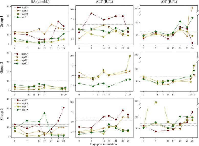
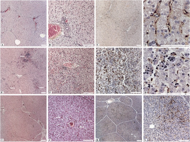
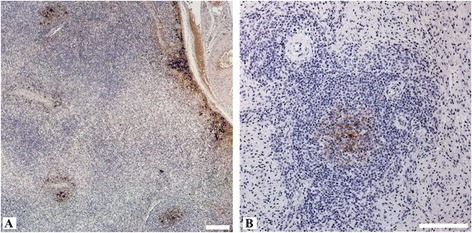
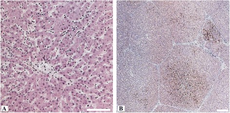
Hepatitis E virus (HEV) is the causative agent of acute hepatitis E in humans in developing countries, but sporadic and autochthonous cases do also occur in industrialised countries. In Europe, food-borne zoonotic transmission of genotype 3 (gt3) has been associated with domestic pig and wild boar. However, little is known about the course of HEV infection in European wild boar and their role in HEV transmission to domestic pigs. To investigate the transmissibility and pathogenesis of wild boar-derived HEVgt3, we inoculated four wild boar and four miniature pigs intravenously. Using quantitative real-time RT-PCR viral RNA was detected in serum, faeces and in liver, spleen and lymph nodes. The antibody response evolved after fourteen days post inoculation. Histopathological findings included mild to moderate lymphoplasmacytic hepatitis which was more prominent in wild boar than in miniature pigs. By immunohistochemical methods, viral antigens were detected mainly in Kupffer cells and liver sinusoidal endothelial cells, partially associated with hepatic lesions, but also in spleen and lymph nodes. While clinical symptoms were subtle and gross pathology was inconspicuous, increased liver enzyme levels in serum indicated hepatocellular injury. As the faecal-oral route is supposed to be the most likely transmission route, we included four contact animals to prove horizontal transmission. Interestingly, HEVgt3-infection was also detected in wild boar and miniature pigs kept in contact to intravenously inoculated wild boar. Given the high virus loads and long duration of viral shedding, wild boar has to be considered as an important HEV reservoir and transmission host in Europe.


Figures

Figure 1
Figure 1
Detection of bile acids (BA), alanine aminotransferase (ALT) and gamma-glutamyl transferase (γGT) in serum. Group 1: Intravenous inoculation of wild boar. Group 2: Intravenous inoculation of miniature pigs. Group 3: Contact infection of wild boar and miniature pigs. For the evaluation of the results, upper reference value ranges for the tested biochemical parameters were calculated. Therefore, different serum samples of the negative control wild boar and miniature pigs were analysed (for each subspecies n =13). Upper reference range limit for wild boar = grey dot-dashed line. Upper reference range limit for miniature pig = grey dot-dot-dashed line. IU = international units. * Sudden death at 1 dpi (after blood collection).
Figure 2
Figure 2
Serology and HEV RNA detection. Group 1: Intravenous inoculation of wild boar. Group 2: Intravenous inoculation of miniature pigs. Group 3: Contact infection of wild boar and miniature pigs. A) Antibody responses to HEV in serum of inoculated wild boar and miniature pigs measured by a double-antigen sandwich ELISA. OD450-values ≥1 are prescribed as seropositive – this threshold is indicated as a grey-dashed line. B) HEV RNA in serum and faeces of HEV inoculated wild boar and miniature pigs estimated by RT-qPCR. * Sudden death at 1 dpi (after blood collection).
Figure 3
Figure 3
Histopathological alterations and immunohistochemistry of the liver from intravenously infected wild boar (Group 1) and miniature pigs (Group 2). A) Hepatic lobules with moderate hyperaemia of sinusoids and portal fields (wb95). B) The lobules show swelling and vacuolation of hepatocytes (wb95). C) Diffuse distribution of viral antigens within the liver lobules (wb95). D) Marked immunolabelling within a hepatic lobule, intracytoplasmatic mainly in Kupffer cells (arrows) and liver sinusoidal endothelial cells (wb93). E) Multifocal hepatocellular degeneration with focus on centrilobular areas (arrows) and hyperaemic central veins (wb11). F) Centrilobular area of hepatocellular degeneration (apoptotic bodies) with infiltrates of lymphocytes, plasma cells and Kupffer cells (wb10). G) Viral antigens within the centrilobular area of a liver lobule in association with degenerated hepatocytes and inflammatory infiltrates (wb11). H) Viral antigens within an area of hepatocellular degeneration, mainly in association with Kupffer cells (arrows) and some hepatocytes (wb10). I) Hepatic lobule with mild hyperaemia of sinusoids and portal fields (mp39). J) Areas of spotty necrosis and apoptotic bodies (arrows) with slight infiltrates of lymphocytes and Kupffer cells (mp37). K) Diffuse distribution of viral antigens within the liver lobules (mp39). L) Intense immunolabelling within a hepatic lobule, mainly in association with Kupffer cells and liver sinusoidal endothelial cells (mp39). All scale bars represent 100 μm.
Figure 4
Figure 4
Immunohistochemistry of liver lymph node and spleen from intravenously HEV inoculated wild boar (Group 1). A) Viral antigens in the subcapsular layer and in the germinal centre of secondary follicles of a liver lymph node (wb93). B) Splenic immunolabelling of viral antigens in the germinal centre of a lymphoid follicle (wb11). All scale bars represent 100 μm.
Figure 5
Figure 5
Histopathological alterations and immunohistochemistry of the liver from the contact wild boar (Group 3). A) Intralobular area with inflammatory infiltrates mostly lymphocytes and histiocytes. B) Multifocal distribution of viral antigens especially in centrilobular areas (arrows). All scale bars represent 100 μm.

References

    1. Tam AW, Smith MM, Guerra ME, Huang CC, Bradley DW, Fry KE, Reyes GR. Hepatitis E virus (HEV): molecular cloning and sequencing of the full-length viral genome. Virology. 1991;185:120–131. doi: 10.1016/0042-6822(91)90760-9. - DOI - PMC - PubMed
    1. Emerson SU, Purcell RH. Hepatitis E virus. Rev Med Virol. 2003;13:145–154. doi: 10.1002/rmv.384. - DOI - PubMed
    1. Emerson SU, Purcell RH. Fields Virology. 2. Philadelphia: Wolters Kluwer Health/Lippincott Williams and Wilkins; 2013. Hepatitis E virus; pp. 2242–2258.
    1. Colson P, Borentain P, Queyriaux B, Kaba M, Moal V, Gallian P, Heyries L, Raoult D, Gerolami R. Pig liver sausage as a source of hepatitis E virus transmission to humans. J Infect Dis. 2010;202:825–834. doi: 10.1086/655898. - DOI - PubMed
    1. Tei S, Kitajima N, Takahashi K, Mishiro S. Zoonotic transmission of hepatitis E virus from deer to human beings. Lancet. 2003;362:371–373. doi: 10.1016/S0140-6736(03)14025-1. - DOI - PubMed

Publication types

LinkOut - more resources