

Novel membrane-based electrochemical sensor for real-time bio-applications
- PMID: 25421738
- PMCID: PMC4279581
- DOI: 10.3390/s141122128
Novel membrane-based electrochemical sensor for real-time bio-applications
Abstract
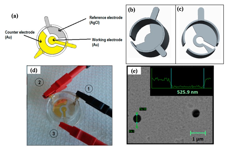
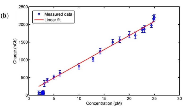
This article presents a novel membrane-based sensor for real-time electrochemical investigations of cellular- or tissue cultures. The membrane sensor enables recording of electrical signals from a cell culture without any signal dilution, thus avoiding loss of sensitivity. Moreover, the porosity of the membrane provides optimal culturing conditions similar to existing culturing techniques allowing more efficient nutrient uptake and molecule release. The patterned sensor electrodes were fabricated on a porous membrane by electron-beam evaporation. The electrochemical performance of the membrane electrodes was characterized by cyclic voltammetry and chronoamperometry, and the detection of synthetic dopamine was demonstrated down to a concentration of 3.1 pM. Furthermore, to present the membrane-sensor functionality the dopamine release from cultured PC12 cells was successfully measured. The PC12 cells culturing experiments showed that the membrane-sensor was suitable as a cell culturing substrate for bio-applications. Real-time measurements of dopamine exocytosis in cell cultures were performed, where the transmitter release was recorded at the point of release. The developed membrane-sensor provides a new functionality to the standard culturing methods, enabling sensitive continuous in vitro monitoring and closely mimicking the in vivo conditions.
Figures


Similar articles
-
Pyrrole-phenylboronic acid: a novel monomer for dopamine recognition and detection based on imprinted electrochemical sensor.Biosens Bioelectron. 2015 Feb 15;64:212-8. doi: 10.1016/j.bios.2014.08.083. Epub 2014 Sep 4. Biosens Bioelectron. 2015. PMID: 25218775
-
Electrochemical sensor based on molecularly imprinted film at polypyrrole-sulfonated graphene/hyaluronic acid-multiwalled carbon nanotubes modified electrode for determination of tryptamine.Biosens Bioelectron. 2012 Jan 15;31(1):277-83. doi: 10.1016/j.bios.2011.10.032. Epub 2011 Oct 24. Biosens Bioelectron. 2012. PMID: 22074810
-
Electrochemical detection of dopamine in the presence of ascorbic acid using graphene modified electrodes.Biosens Bioelectron. 2010 Jun 15;25(10):2366-9. doi: 10.1016/j.bios.2010.02.031. Epub 2010 Mar 4. Biosens Bioelectron. 2010. PMID: 20307965
-
Polymer thin films embedded with metal nanoparticles for electrochemical biosensors applications.Biosens Bioelectron. 2013 Mar 15;41:43-53. doi: 10.1016/j.bios.2012.09.031. Epub 2012 Sep 29. Biosens Bioelectron. 2013. PMID: 23083910 Review.
-
Silicon nanowire field-effect-transistor based biosensors: from sensitive to ultra-sensitive.Biosens Bioelectron. 2014 Oct 15;60:101-11. doi: 10.1016/j.bios.2014.03.057. Epub 2014 Apr 15. Biosens Bioelectron. 2014. PMID: 24787124 Review.
Cited by
-
Gold Electrodes Modified with Calix[4]arene for Electrochemical Determination of Dopamine in the Presence of Selected Neurotransmitters.Sensors (Basel). 2017 Jun 13;17(6):1368. doi: 10.3390/s17061368. Sensors (Basel). 2017. PMID: 28608815 Free PMC article.
-
Evolvable Smartphone-Based Platforms for Point-of-Care In-Vitro Diagnostics Applications.Diagnostics (Basel). 2016 Sep 3;6(3):33. doi: 10.3390/diagnostics6030033. Diagnostics (Basel). 2016. PMID: 27598208 Free PMC article.
-
Fast Selective Detection of Pyocyanin Using Cyclic Voltammetry.Sensors (Basel). 2016 Mar 19;16(3):408. doi: 10.3390/s16030408. Sensors (Basel). 2016. PMID: 27007376 Free PMC article.
-
Development of biosensors for detection of fibrinogen: a review.Anal Bioanal Chem. 2024 Jan;416(1):21-36. doi: 10.1007/s00216-023-04976-1. Epub 2023 Oct 14. Anal Bioanal Chem. 2024. PMID: 37837539 Review.
-
PC-12 Cell Line as a Neuronal Cell Model for Biosensing Applications.Biosensors (Basel). 2022 Jul 8;12(7):500. doi: 10.3390/bios12070500. Biosensors (Basel). 2022. PMID: 35884303 Free PMC article. Review.
References
-
- Wang J. Modified electrodes for electrochemical sensors. Electroanalysis. 1990;3:255–259.
-
- Nigovic B., Spajic J. A novel electrochemical sensor for assaying of antiphyshotic drug quetiapine. Talanta. 2011;86:393–399. - PubMed
-
- Castillo J.J., Svendsen E.W., Rozlosnik N., Escobar P., Martineza F., Castillo-León J. Detection of cancer cells using a peptide nanotube-folic acid modified graphene electrode. Analyst. 2013;138:1026–1031. - PubMed
-
- Drummond T.G., Hill M.G. Electrochemical DNA sensors. Nat. Biotechnol. 2003;21:1192–1199. - PubMed
MeSH terms
Substances
LinkOut - more resources
Full Text Sources
Other Literature Sources
