Review
. 2014 Nov 21;6(11):4536-70.
doi: 10.3390/v6114536.

Structure, function and dynamics in adenovirus maturation

Affiliations
Review

Structure, function and dynamics in adenovirus maturation

Walter F Mangel et al. Viruses. .

Abstract

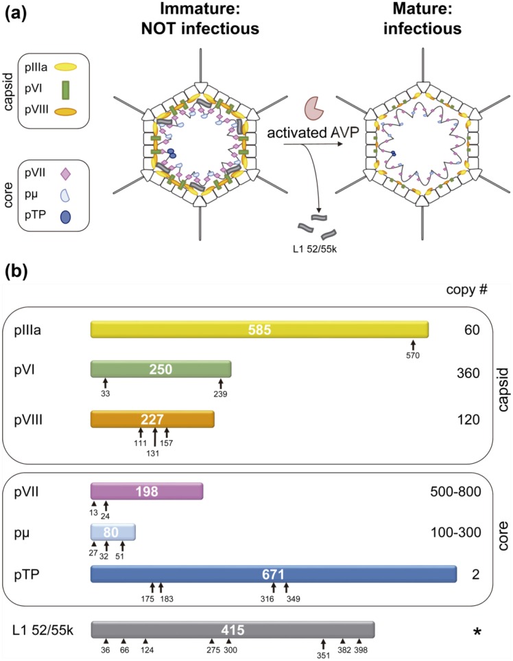
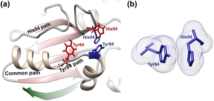
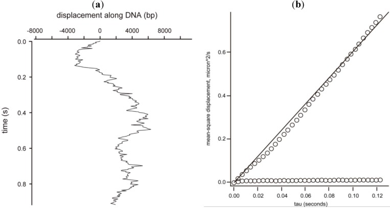
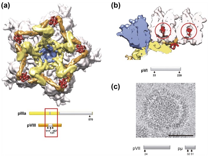
Here we review the current knowledge on maturation of adenovirus, a non-enveloped icosahedral eukaryotic virus. The adenovirus dsDNA genome fills the capsid in complex with a large amount of histone-like viral proteins, forming the core. Maturation involves proteolytic cleavage of several capsid and core precursor proteins by the viral protease (AVP). AVP uses a peptide cleaved from one of its targets as a "molecular sled" to slide on the viral genome and reach its substrates, in a remarkable example of one-dimensional chemistry. Immature adenovirus containing the precursor proteins lacks infectivity because of its inability to uncoat. The immature core is more compact and stable than the mature one, due to the condensing action of unprocessed core polypeptides; shell precursors underpin the vertex region and the connections between capsid and core. Maturation makes the virion metastable, priming it for stepwise uncoating by facilitating vertex release and loosening the condensed genome and its attachment to the icosahedral shell. The packaging scaffold protein L1 52/55k is also a substrate for AVP. Proteolytic processing of L1 52/55k disrupts its interactions with other virion components, providing a mechanism for its removal during maturation. Finally, possible roles for maturation of the terminal protein are discussed.


Figures

Figure 1
Figure 1
Substrates of the AdV maturation protease, AVP. (a) Schematics showing the location of substrates in the viral particle. The internal location of L1 52/55k is inferred from its interactions with core elements [59]; (b) Each HAdV-C2 precursor protein is represented as a bar with the polypeptide length in amino acids indicated in the center. Consensus cleavage sites are denoted by arrows, reported non-consensus sites by arrowheads [53,60]. The prefix “p” denotes the unprocessed precursors. Copy numbers are derived from stoichiometric analyses [18,19]. A star (*) in place of the copy number for L1 52/55k indicates that its copy number varies depending on the assembly stage: 100 copies in empty particles, 50 in fully packaged, immature ts1 particles, and 0 in mature virions [46,60]. Panel (b) modified from [7].
Figure 2
Figure 2
Conservation of AVP cleavage sites in the L1 52/55k protein. (a) Conservation across human AdV species; (b) Conservation across mastadenovirus species. Sequences were downloaded from GenBank and aligned with TCoffee [70]. The figure was created using JalView [71]. HAdV: human adenovirus; BAdV: bovine adenovirus; CAdV: canine adenovirus; EqAdV: equine adenovirus; SAdV: simian adenovirus; MuAdV: murine adenovirus; OAdV: ovine adenovirus; PAdV: porcine adenovirus; TsAdV: tree shrew adenovirus; BatAdV: bat adenovirus. Black frames indicate the AVP consensus cleavage site in HAdV-C2 L1 52/55k; gray frames indicate non-consensus sites described in [60]; red ovals indicate Gly stretches that could be possible additional cleavage sites.
Figure 2
Figure 2
Conservation of AVP cleavage sites in the L1 52/55k protein. (a) Conservation across human AdV species; (b) Conservation across mastadenovirus species. Sequences were downloaded from GenBank and aligned with TCoffee [70]. The figure was created using JalView [71]. HAdV: human adenovirus; BAdV: bovine adenovirus; CAdV: canine adenovirus; EqAdV: equine adenovirus; SAdV: simian adenovirus; MuAdV: murine adenovirus; OAdV: ovine adenovirus; PAdV: porcine adenovirus; TsAdV: tree shrew adenovirus; BatAdV: bat adenovirus. Black frames indicate the AVP consensus cleavage site in HAdV-C2 L1 52/55k; gray frames indicate non-consensus sites described in [60]; red ovals indicate Gly stretches that could be possible additional cleavage sites.
Figure 3
Figure 3
Crystal structure of the AVP-pVIc complex and locations of the four amino acid residues involved in catalysis in AVP and in AVP-pVIc. (a) Secondary structure of the AVP-pVIc complex with the four amino acid residues involved in catalysis in blue and the pVIc peptide in green; (b) The four amino acids involved in catalysis in AVP-pVIc (blue) and in AVP (red) are juxtaposed. Only His54 is in a different position in the two structures. Figure created with UCSF Chimera (http://www.cgl.ucsf.edu/chimera/) [90].
Figure 4
Figure 4
AVP activation pathway and cation-π interaction. (a) Upon binding of pVIc (green) to AVP, a series of contiguous conformational changes occur along a common path that bifurcates into an upper and lower path. At the end of the upper path, His54 drops down from its position in AVP (red) to a position in AVP-pVIc (blue) that is opposite Cys122. At the end of the lower path, Tyr84 (red) moves 11 Å to a position in AVP-pVIc (blue) where it can form a cation-π interaction with His54; (b) Electron clouds of Tyr84 and His54 in their cation-π interaction. Figure created with UCSF Chimera [90].
Figure 5
Figure 5
Sliding of the AVP-pVIc complex along DNA. (a) An AVP-pVIc complex sliding almost 16,000 bp in less than 1 s; (b) Mean square displacement (MSD) versus time of the data in (a). The MSD is the square of the distance traveled. The slope of the curve is the one-dimensional diffusion constant for this slide, 32 × 106 bp2/s. The line parallel to the abscissa is the MSD from the Y-axis versus time.
Figure 6
Figure 6
Structural differences between immature (ts1) and mature (wt) AdV virions. (a) View from inside the capsid looking at the 5-fold icosahedral symmetry axis, with the density for the molecular stitch derived from the ts1-wt difference map at 8.9 Å resolution in red [128]. The five peripentonal hexons are shown in pale pink; penton base in blue; polypeptide IIIa in yellow; and polypeptide VIII in tan. Surfaces in (a) and (b) were created from the HAdV-C5 high resolution cryo-EM structure (PDB ID 3IYN) [8] and represented with UCSF Chimera [90]. The bars represent the precursor polypeptides IIIa and VIII with the cleavage sites indicated (arrows). Polypeptide regions not traced in the cryo-EM HAdV-C5 high resolution structure are in gray. Untraced regions close to the molecular stitch are indicated with a red rectangle. Modified from [7]; (b) A section across the capsid showing the density attributed to the precursor of protein VI (red circles) inside the inner cavities of two hexon trimers [128]. Colors are as those shown in (a). Density for the molecular stitch is also seen in this view, wedged between polypeptide IIIa and VIII. The bar represents the precursor polypeptide VI with the cleavage sites indicated (arrows); (c) A disrupted particle found in a cryo-EM preparation of ts1 virus, showing the capsid separating from the core, while the latter remains as a compact sphere. The bar in the micrograph represents 50 nm. The bars below represent the precursor polypeptides pVII and pμ with the cleavage sites indicated (arrows). Reproduced with permission from Reference [128]. Copyright 2009, Elsevier.

References

    1. Berk A.J. Adenoviridae: The viruses and their replication. In: Knipe D.M., Howley P.M., Griffin D.E., Lamb R.A., Martin M.A., editors. Fields Virology. 5th ed. Lippincott Williams & Wilkins; Philadelphia, PA, USA: 2007. pp. 2355–2394.
    1. Harrach B., Benkő M., Both G., Brown M., Davison A., Echavarría M., Hess M., Jones M., Kajon A., Lehmkuhl H., et al. In: Virus Taxonomy: Classification and Nomenclature of Viruses. Ninth Report of the International Committee on Taxonomy of Viruses. King A., Adams M., Carstens E., Lefkowitz E., editors. Elsevier; San Diego, CA, USA: 2011. pp. 95–111.
    1. Lasaro M.O., Ertl H.C. New insights on adenovirus as vaccine vectors. Mol. Ther. 2009;17:1333–1339. doi: 10.1038/mt.2009.130. - DOI - PMC - PubMed
    1. Gonçalves M.A.F.V., de Vries A.A.F. Adenovirus: From foe to friend. Rev. Med. Virol. 2006;16:167–186. doi: 10.1002/rmv.494. - DOI - PubMed
    1. Yamamoto M., Curiel D.T. Current issues and future directions of oncolytic adenoviruses. Mol. Ther. 2010;18:243–250. doi: 10.1038/mt.2009.266. - DOI - PMC - PubMed

Publication types