Review
. 2014 Nov 24;6(11):4683-702.
doi: 10.3390/v6114683.

Hepatitis B virus HBx protein interactions with the ubiquitin proteasome system

Affiliations
Review

Hepatitis B virus HBx protein interactions with the ubiquitin proteasome system

Marissa M Minor et al. Viruses. .

Abstract

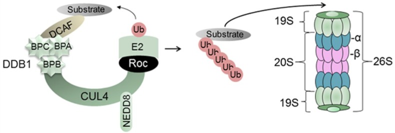
The hepatitis B virus (HBV) causes acute and chronic hepatitis, and the latter is a major risk factor for the development of hepatocellular carcinoma (HCC). HBV encodes a 17-kDa regulatory protein, HBx, which is required for virus replication. Although the precise contribution(s) of HBx to virus replication is unknown, many viruses target cellular pathways to create an environment favorable for virus replication. The ubiquitin proteasome system (UPS) is a major conserved cellular pathway that controls several critical processes in the cell by regulating the levels of proteins involved in cell cycle, DNA repair, innate immunity, and other processes. We summarize here the interactions of HBx with components of the UPS, including the CUL4 adaptor DDB1, the cullin regulatory complex CSN, and the 26S proteasome. Understanding how these protein interactions benefit virus replication remains a challenge due to limited models in which to study HBV replication. However, studies from other viral systems that similarly target the UPS provide insight into possible strategies used by HBV.


Figures

Figure 1
Figure 1
Schematic of the UPS. In the example provided, the E3 ligase consists of the CUL4 scaffold, the DDB1 adaptor protein (folded into three β-propellar, or BP, domains), and the RING protein (Roc) that binds to the E2 enzyme. This complex recruits substrate proteins for ubiquitination and degradation by the 26S proteasome, as described in the text.
Figure 2
Figure 2
HBV genome organization. HBV contains a small, partially double-stranded (~dsDNA) genome (see inner black circles) that consists of a full-length negative strand and an incomplete (dashed lines) positive strand. The genome contains four promoters, two enhancer regions (Enh1, Enh2), and two direct repeats (DR1, DR2). The four ORFs are depicted by the colored arrows. During virus replication, the ~dsDNA genome is repaired into covalently closed circular (ccc) DNA, which serves as the template for viral transcription. The four major transcripts, shown as thin black outer arrows, are described in the text. Note that the X ORF (red) is present in all four HBV mRNAs.
Figure 3
Figure 3
Schematic representation of HBx. The 154-aa HBx protein is conserved in all mammalian hepadnaviruses. Functional domains shown include the regulatory domain (aa 1–50), the transactivation domain (aa 50–154), and the DDB1 binding domain (aa 88–100; hatched area). The proteasome binding domain (aa 90–154) spans the DDB1 binding domain [69]. Conserved cysteine residues are shown by the asterisks (aa 7, 17, 61, 69, 78, 115, 137, 143, and 148) [70] and have an unknown function. Six conserved lysine residues, which may be targets for ubiquitination, are indicated as filled triangles (aa 91, 95, 113, 118, 130, 140) [70,71].
Figure 4
Figure 4
DDB1 binding motif found in DCAFs. (A), A DDB1-WD40 box (DWD) domain is present in some WD40-repeat-containing proteins [19] and mediates the binding of these DCAFs to DDB1. The 16-aa motif, estimated to occur in 79 genes in humans, is based on the biochemical/biophysical properties of the aa. (B), HBx sequences from three HBV subtypes show a similar DWD motif. Reproduced with permission from Reference [78]. Copyright 2008, Research Signpost.

References

    1. Pickart C.M. Back to the future with ubiquitin. Cell. 2004;116:181–190. doi: 10.1016/S0092-8674(03)01074-2. - DOI - PubMed
    1. Voutsadakis I.A. Ubiquitin, ubiquitination and the ubiquitin-proteasome system in cancer. Atlas Genet. Cytogenet. Oncol. Haematol. 2010;14:1088–1099.
    1. Brooks P., Fuertes G., Murray R.Z., Bose S., Knecht E., Rechsteiner M.C., Hendil K.B., Tanaka K., Dyson J., Rivett J. Subcellular localization of proteasomes and their regulatory complexes in mammalian cells. Biochem. J. 2000;346:155–161. doi: 10.1042/0264-6021:3460155. - DOI - PMC - PubMed
    1. Petroski M.D., Deshaies R.J. Function and regulation of Cullin-RING ubiquitin ligases. Nat. Rev. Mol. Cell Biol. 2005;6:9–20. doi: 10.1038/nrm1547. - DOI - PubMed
    1. Gustin J.K., Moses A.V., Fruh K., Douglas J.L. Viral takeover of the host ubiquitin system. Front. Microbiol. 2011;2:161. doi: 10.3389/fmicb.2011.00161. - DOI - PMC - PubMed

Publication types