. 2014 Dec 15;211(13):2487-96.
doi: 10.1084/jem.20141788. Epub 2014 Nov 24.

Potential role of orexin and sleep modulation in the pathogenesis of Alzheimer's disease

Affiliations

Potential role of orexin and sleep modulation in the pathogenesis of Alzheimer's disease

Jee Hoon Roh et al. J Exp Med. .

Erratum in

  • J Exp Med. 2015 Jan 12;212(1):121

Abstract

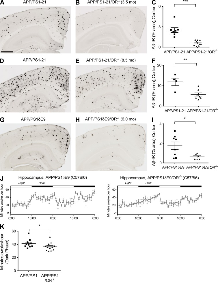
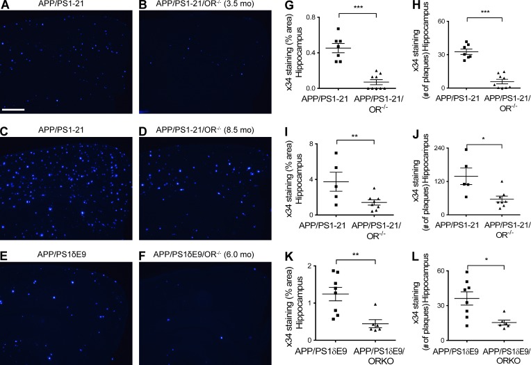
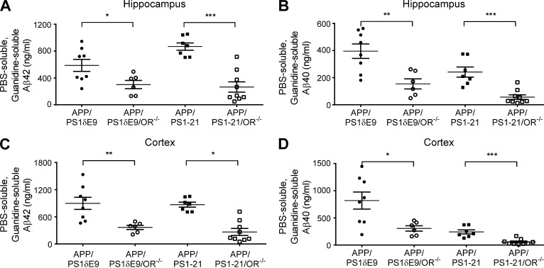
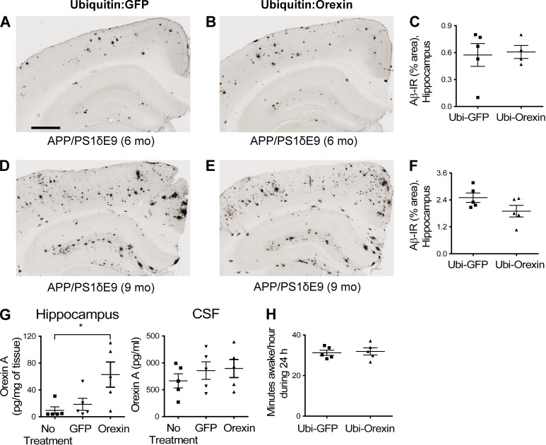
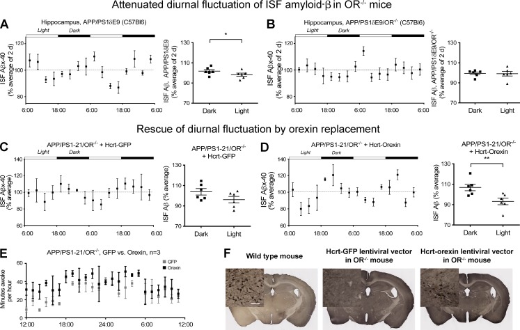
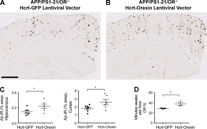
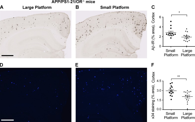
Age-related aggregation of amyloid-β (Aβ) is an upstream pathological event in Alzheimer's disease (AD) pathogenesis, and it disrupts the sleep-wake cycle. The amount of sleep declines with aging and to a greater extent in AD. Poor sleep quality and insufficient amounts of sleep have been noted in humans with preclinical evidence of AD. However, how the amount and quality of sleep affects Aβ aggregation is not yet well understood. Orexins (hypocretins) initiate and maintain wakefulness, and loss of orexin-producing neurons causes narcolepsy. We tried to determine whether orexin release or secondary changes in sleep via orexin modulation affect Aβ pathology. Amyloid precursor protein (APP)/Presenilin 1 (PS1) transgenic mice, in which the orexin gene is knocked out, showed a marked decrease in the amount of Aβ pathology in the brain with an increase in sleep time. Focal overexpression of orexin in the hippocampus in APP/PS1 mice did not alter the total amount of sleep/wakefulness and the amount of Aβ pathology. In contrast, sleep deprivation or increasing wakefulness by rescue of orexinergic neurons in APP/PS1 mice lacking orexin increased the amount of Aβ pathology in the brain. Collectively, modulation of orexin and its effects on sleep appear to modulate Aβ pathology in the brain.


Figures

Figure 1.
Figure 1.
Marked reduction of Aβ pathology in the APP/PS1/OR−/− mice compared with APP/PS1 mice. (A–I) The amount of Aβ pathology was noted at 3.5 (A–C) and 8.5 mo (D–F) in APP/PS1-21 mouse line and at 6 mo in APP/PS1δE9 mouse line (G–I). (J and K) Amount of wakefulness at 3 mo of age before the onset of Aβ pathology in the brain was compared between APP/PS1δE9 and APP/PS1δE9/OR−/− mice. Each mouse was investigated independently one time. Data are presented as mean ± SEM (n = 4–12 in each group, two-tailed Student’s t test in C, F, and I and Mann-Whitney test in J). *, P < 0.05; **, P < 0.01; and ***, P < 0.001. Bar, 500 µm.
Figure 2.
Figure 2.
Marked reduction of amyloid pathology in the APP/PS1/OR−/− mice compared with APP/PS1 mice. (A–L) Amyloid pathology measured by Aβ immunoreactivity and number of amyloid plaques after X-34 staining is noted at 3.5 (A, B, G, and H) and 8.5 mo (C, D, I, and J) in the APP/PS1-21 mouse line and at 6 mo in the APP/PS1δE9 mouse line (E, F, K, and L). Each mouse was investigated independently one time. Data are presented as mean ± SEM (n = 5–9 in each group, two-tailed Student’s t test). *, P < 0.05; **, P < 0.01; and ***, P < 0.001. Bar, 100 µm.
Figure 3.
Figure 3.
Strong reduction in Aβ species in APP/PS1 mice lacking orexin. (A–D) The amount of PBS-soluble and guanidine-soluble forms of Aβ40 and Aβ42 were compared in APP/PS1δE9/OR−/− and APP/PS1δE9 mice (left two columns) and in APP/PS1-21/OR−/− and APP/PS1-21 mice (right two columns). Results were obtained from the hippocampus (A and B) and from the cortex (C and D) of each group of mice. Each mouse was investigated independently one time. All samples were measured in triplicate. Data are presented as mean ± SEM (n = 5–9 in each group, two-tailed Student’s t test). *, P < 0.05; **, P < 0.01; and ***, P < 0.001.
Figure 4.
Figure 4.
No changes in Aβ deposition and amount of wakefulness by focal overexpression of orexin. (A–F) Amount of Aβ pathology was compared after focal injection of ubiquitin-driven orexin lentiviral vector versus ubiquitin-driven GFP lentiviral vector in the hippocampus from 3 to 6 mo (A–C) or 5 to 9 mo (D–F) in APP/PS1δE9 mice. (G) Levels of orexin in the hippocampus and CSF were compared after focal injection of orexin or GFP lentiviral vector driven by ubiquitin promoter. (H) The amount of wakefulness in APP/PS1δE9 mice was compared after focal injection of ubiquitin-driven orexin lentiviral vector versus ubiquitin-driven GFP lentiviral vector. Each mouse was investigated independently one time. Data are presented as mean ± SEM (n = 4–5 in each group, two-tailed Student’s t test in C, F, and H; one-way ANOVA, followed by Tukey’s post hoc test in G). *, P < 0.05. Bar, 500 µm.
Figure 5.
Figure 5.
Restoration of diurnal fluctuation of ISF Aβ40 via rescue of orexin expression in APP/PS1/OR−/− mice. (A and B) Diurnal fluctuation of ISF Aβ40 in young APP/PS1δE9 mice was compared with the diurnal fluctuation in the same mice when orexin was knocked out (APP/PS1δE9/OR−/− mice). (C–E) Diurnal fluctuation of ISF Aβ40 in the hippocampus and minutes awake per hour of APP/PS1-21/OR−/− mice treated with hypocretin/orexin promoter–driven lentiviral vector overexpressing orexin in the hypothalamus bilaterally were compared with mice treated with the same lentiviral vector expressing GFP. (F) Expression of orexin by immunostaining in orexinergic neurons in the hypothalamus of APP/PS1-21/OR−/− mice treated with hypocretin/orexin promoter–driven lentiviral vector expressing GFP or orexin (middle and right) compared with wild-type mice (left). Each mouse was investigated independently one time. All ISF Aβ measurements were performed in duplicate. Data are presented as mean ± SEM (n = 6 in each group, two-tailed Student’s t test in A–D and Mann–Whitney test in E). *, P < 0.05; and **, P < 0.01. Bar, 100 µm.
Figure 6.
Figure 6.
Increase in Aβ deposition and wakefulness by rescue of orexin expression in APP/PS1/OR−/− mice via expression of orexin with lentiviral vector driven by hypocretin/orexin promoter in the bilateral hypothalamus. (A–C) Amount of Aβ pathology in the hippocampus and cortex of the APP/PS1-21/OR−/− mice after bilateral injection of hypocretin promoter–driven orexin lentiviral vector in the hypothalamus of the APP/PS1/OR−/− mice was compared with results from injection of the same lentiviral vector overexpressing GFP. (D) The amount of wakefulness as measured by polysomnography was compared between the same groups of mice. Each mouse was investigated independently one time. Data are presented as mean ± SEM (n = 6–7 in each group, two-tailed Student’s t test in C; n = 3 in each group, Mann–Whitney test in D). *, P < 0.05. Bar, 500 µm.
Figure 7.
Figure 7.
Increased Aβ deposition with chronic sleep deprivation in APP/PS1-21/OR−/− mice. (A–F) The amount of Aβ plaques stained with HJ3.4B (A–C) and fibrillar Aβ stained with X-34 (D–F) were compared between APP/PS1-21/OR−/− mice exposed to a large platform and a small platform. Sleep deprivation experiments were performed using a small and large platform in a cage with water on the bottom, where a mouse cannot sleep on the small platform because of its size, whereas they can maintain a normal sleep–wake cycle on the larger platform. Mice exposed to small platforms (n = 3) and to large platforms (n = 3) were analyzed together within a set of experiments. The results are the sum of five repeats in different mice. Each mouse was investigated independently. Data are presented as mean ± SEM (n = 13–14 in each group, two-tailed Student’s t test). *, P < 0.05; and **, P < 0.01. Bars: (A) 500 µm; (D) 100 µm.

Comment in

  • Sleeping our Alzheimer's risk away.
    Watts RJ. Watts RJ. J Exp Med. 2014 Dec 15;211(13):2484-5. doi: 10.1084/jem.21113insight4. J Exp Med. 2014. PMID: 25512582 Free PMC article. No abstract available.

References

    1. Bero A.W., Yan P., Roh J.H., Cirrito J.R., Stewart F.R., Raichle M.E., Lee J.M., and Holtzman D.M.. 2011. Neuronal activity regulates the regional vulnerability to amyloid-β deposition. Nat. Neurosci. 14:750–756 10.1038/nn.2801 - DOI - PMC - PubMed
    1. Carter M.E., Yizhar O., Chikahisa S., Nguyen H., Adamantidis A., Nishino S., Deisseroth K., and de Lecea L.. 2010. Tuning arousal with optogenetic modulation of locus coeruleus neurons. Nat. Neurosci. 13:1526–1533 10.1038/nn.2682 - DOI - PMC - PubMed
    1. Chemelli R.M., Willie J.T., Sinton C.M., Elmquist J.K., Scammell T., Lee C., Richardson J.A., Williams S.C., Xiong Y., Kisanuki Y., et al. . 1999. Narcolepsy in orexin knockout mice: molecular genetics of sleep regulation. Cell. 98:437–451 10.1016/S0092-8674(00)81973-X - DOI - PubMed
    1. Cirrito J.R., May P.C., O’Dell M.A., Taylor J.W., Parsadanian M., Cramer J.W., Audia J.E., Nissen J.S., Bales K.R., Paul S.M., et al. . 2003. In vivo assessment of brain interstitial fluid with microdialysis reveals plaque-associated changes in amyloid-β metabolism and half-life. J. Neurosci. 23:8844–8853. - PMC - PubMed
    1. Cirrito J.R., Kang J.E., Lee J., Stewart F.R., Verges D.K., Silverio L.M., Bu G., Mennerick S., and Holtzman D.M.. 2008. Endocytosis is required for synaptic activity-dependent release of amyloid-β in vivo. Neuron. 58:42–51 10.1016/j.neuron.2008.02.003 - DOI - PMC - PubMed

Publication types

MeSH terms