

Adenosine monophosphate-activated protein kinase activation enhances embryonic neural stem cell apoptosis in a mouse model of amyotrophic lateral sclerosis
- PMID: 25422638
- PMCID: PMC4238165
- DOI: 10.4103/1673-5374.143421
Adenosine monophosphate-activated protein kinase activation enhances embryonic neural stem cell apoptosis in a mouse model of amyotrophic lateral sclerosis
Abstract
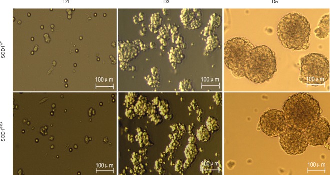
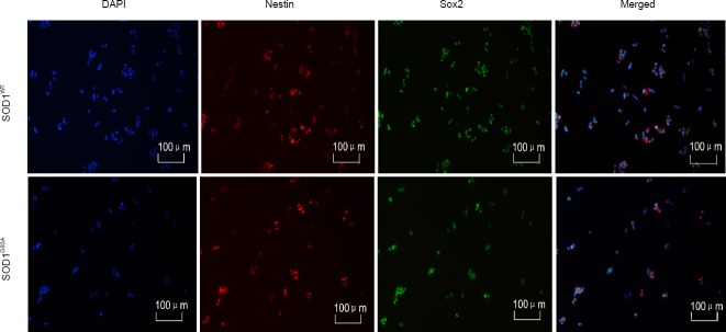
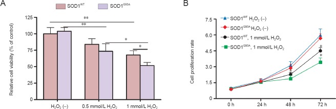
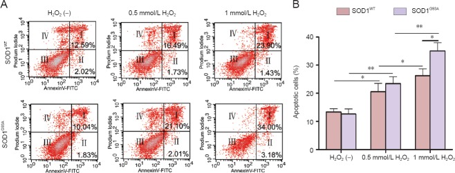
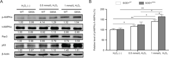
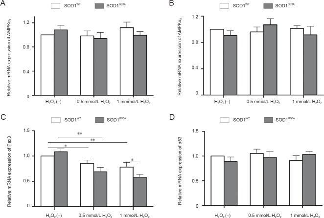
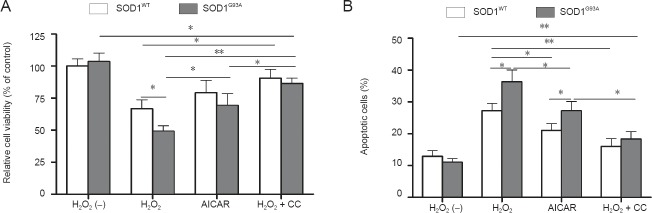
Alterations in embryonic neural stem cells play crucial roles in the pathogenesis of amyotrophic lateral sclerosis. We hypothesized that embryonic neural stem cells from SOD1(G93A) individuals might be more susceptible to oxidative injury, resulting in a propensity for neurodegeneration at later stages. In this study, embryonic neural stem cells obtained from human superoxide dismutase 1 mutant (SOD1(G93A)) and wild-type (SOD1(WT)) mouse models were exposed to H2O2. We assayed cell viability with mitochondrial succinic dehydrogenase colorimetric reagent, and measured cell apoptosis by flow cytometry. Moreover, we evaluated the expression of the adenosine monophosphate-activated protein kinase (AMPK) α-subunit, paired box 3 (Pax3) protein, and p53 in western blot analyses. Compared with SOD1(WT) cells, SOD1(G93A) embryonic neural stem cells were more likely to undergo H2O2-induced apoptosis. Phosphorylation of AMPKα in SOD1(G93A) cells was higher than that in SOD1(WT) cells. Pax3 expression was inversely correlated with the phosphorylation levels of AMPKα. p53 protein levels were also correlated with AMPKα phosphorylation levels. Compound C, an inhibitor of AMPKα, attenuated the effects of H2O2. These results suggest that embryonic neural stem cells from SOD1(G93A) mice are more susceptible to apoptosis in the presence of oxidative stress compared with those from wild-type controls, and the effects are mainly mediated by Pax3 and p53 in the AMPKα pathway.
Keywords: NSFC grants; SOD1G93A mouse; adenosine monophosphate-activated protein kinase α; amyotrophic lateral sclerosis; apoptosis; embryonic neural stem cells; hydrogen peroxide; nerve regeneration; neural regeneration; neuroderegeneration; oxidative stress; p53; paired box 3.
Conflict of interest statement
Figures

Similar articles
-
Effects of diet on adenosine monophosphate-activated protein kinase activity and disease progression in an amyotrophic lateral sclerosis model.J Int Med Res. 2015 Feb;43(1):67-79. doi: 10.1177/0300060514554725. Epub 2014 Dec 22. J Int Med Res. 2015. PMID: 25534414
-
Hypoxia causes autophagic stress and derangement of metabolic adaptation in a cell model of amyotrophic lateral sclerosis.J Neurochem. 2014 May;129(3):413-25. doi: 10.1111/jnc.12642. Epub 2014 Jan 22. J Neurochem. 2014. PMID: 24359187
-
"Preconditioning" with latrepirdine, an adenosine 5'-monophosphate-activated protein kinase activator, delays amyotrophic lateral sclerosis progression in SOD1(G93A) mice.Neurobiol Aging. 2015 Feb;36(2):1140-50. doi: 10.1016/j.neurobiolaging.2014.09.022. Epub 2014 Sep 26. Neurobiol Aging. 2015. PMID: 25443289
-
Progressive Degeneration and Inhibition of Peripheral Nerve Regeneration in the SOD1-G93A Mouse Model of Amyotrophic Lateral Sclerosis.Cell Physiol Biochem. 2018;46(6):2358-2372. doi: 10.1159/000489627. Epub 2018 May 4. Cell Physiol Biochem. 2018. PMID: 29742495
-
On the relation of oxidative stress to neuroinflammation: lessons learned from the G93A-SOD1 mouse model of amyotrophic lateral sclerosis.Antioxid Redox Signal. 2006 Nov-Dec;8(11-12):2075-87. doi: 10.1089/ars.2006.8.2075. Antioxid Redox Signal. 2006. PMID: 17034351 Review.
Cited by
-
Sephin1 suppresses ER stress-induced cell death by inhibiting the formation of PP2A holoenzyme.Cell Death Dis. 2025 Feb 19;16(1):117. doi: 10.1038/s41419-025-07450-1. Cell Death Dis. 2025. PMID: 39971896 Free PMC article.
-
Metabolism Regulation and Redox State: Insight into the Role of Superoxide Dismutase 1.Int J Mol Sci. 2020 Sep 10;21(18):6606. doi: 10.3390/ijms21186606. Int J Mol Sci. 2020. PMID: 32927603 Free PMC article. Review.
-
Cysteine Modifications in the Pathogenesis of ALS.Front Mol Neurosci. 2017 Jan 23;10:5. doi: 10.3389/fnmol.2017.00005. eCollection 2017. Front Mol Neurosci. 2017. PMID: 28167899 Free PMC article. Review.
-
Exploring the Role of Licorice and Its Derivatives in Cell Signaling Pathway NF-κB and MAPK.J Nutr Metab. 2024 Oct 23;2024:9988167. doi: 10.1155/2024/9988167. eCollection 2024. J Nutr Metab. 2024. PMID: 39479405 Free PMC article. Review.
-
Protein kinases in neurodegenerative diseases: current understandings and implications for drug discovery.Signal Transduct Target Ther. 2025 May 7;10(1):146. doi: 10.1038/s41392-025-02179-x. Signal Transduct Target Ther. 2025. PMID: 40328798 Free PMC article. Review.
References
-
- Beleza-Meireles A, Al-Chalabi A. Genetic studies of amyotrophic lateral sclerosis: controversies and perspectives. Amyotroph Lateral Scler. 2009;10:1–14. - PubMed
-
- Bouteloup C, Desport JC, Clavelou P, Guy N, Derumeaux-Burel H, Ferrier A, Couratier P. Hypermetabolism in ALS patients: an early and persistent phenomenon. J Neurol. 2009;256:1236–1242. - PubMed
-
- Bruijn LI, Becher MW, Lee MK, Anderson KL, Jenkins NA, Copeland NG, Sisodia SS, Rothstein JD, Borchelt DR, Price DL, Cleveland DW. ALS-linked SOD1 mutant G85R mediates damage to astrocytes and promotes rapidly progressive disease with SOD1-containing inclusions. Neuron. 1997;18:327–338. - PubMed
-
- Cai B, Fan DS. Germline degradation of a mouse model of familial amyotrophic lateral sclerosis when breeding. Zhongguo Zuzhi Gongcheng Yanjiu. 2013;17:4521–4528.
LinkOut - more resources
Full Text Sources
Other Literature Sources
Research Materials
Miscellaneous