

Function of microglia and macrophages in secondary damage after spinal cord injury
- PMID: 25422640
- PMCID: PMC4239768
- DOI: 10.4103/1673-5374.143423
Function of microglia and macrophages in secondary damage after spinal cord injury
Abstract
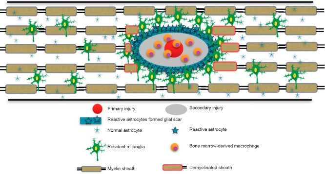
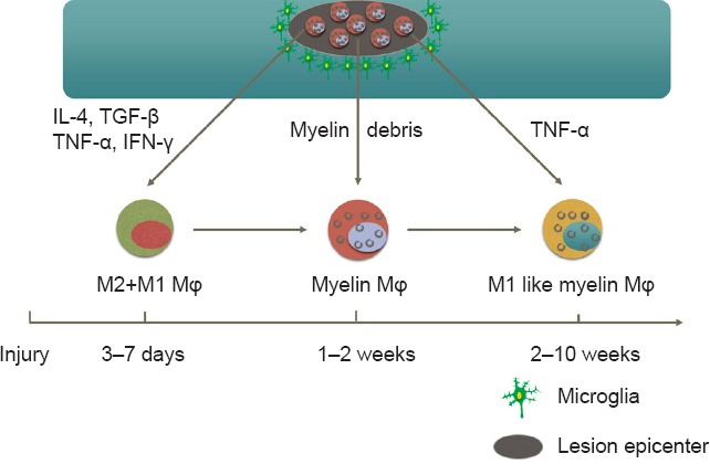
Spinal cord injury (SCI) is a devastating type of neurological trauma with limited therapeutic opportunities. The pathophysiology of SCI involves primary and secondary mechanisms of injury. Among all the secondary injury mechanisms, the inflammatory response is the major contributor and results in expansion of the lesion and further loss of neurologic function. Meanwhile, the inflammation directly and indirectly dominates the outcomes of SCI, including not only pain and motor dysfunction, but also preventingneuronal regeneration. Microglia and macrophages play very important roles in secondary injury. Microglia reside in spinal parenchyma and survey the microenvironment through the signals of injury or infection. Macrophages are derived from monocytes recruited to injured sites from the peripheral circulation. Activated resident microglia and monocyte-derived macrophages induce and magnify immune and inflammatory responses not only by means of their secretory moleculesand phagocytosis, but also through their influence on astrocytes, oligodendrocytes and demyelination. In this review, we focus on the roles of microglia and macrophages in secondary injury and how they contribute to the sequelae of SCI.
Keywords: M1/M2 activation; astrocytes; chemokines; cytokines; demyelination; inflammation; macrophages; microglia; oligodendrocytes; secondary damage; spinal cord injury.
Figures
References
-
- Ahn M, Lee C, Jung K, Kim H, Moon C, Sim KB, Shin T. Immunohistochemical study of arginase-1 in the spinal cords of rats with clip compression injury. Brain Res. 2012;1445:11–19. - PubMed
-
- Beuche W, Friede RL. The role of non-resident cells in wallerian degeneration. J Neurocytol. 1984;13:767–796. - PubMed
Publication types
Grants and funding
LinkOut - more resources
Full Text Sources
Other Literature Sources