

doi: 10.1038/nmeth.3175.
Live-cell mass profiling: an emerging approach in quantitative biophysics
Affiliations
- PMID: 25423019
- PMCID: PMC4319180
- DOI: 10.1038/nmeth.3175
Item in Clipboard
Live-cell mass profiling: an emerging approach in quantitative biophysics
Nat Methods.
2014 Dec.
Abstract
Cell mass, volume and growth rate are tightly controlled biophysical parameters in cellular development and homeostasis, and pathological cell growth defines cancer in metazoans. The first measurements of cell mass were made in the 1950s, but only recently have advances in computer science and microfabrication spurred the rapid development of precision mass-quantifying approaches. Here we discuss available techniques for quantifying the mass of single live cells with an emphasis on relative features, capabilities and drawbacks for different applications.
Figures
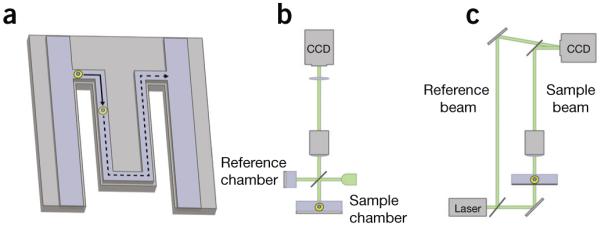
Some methods for single-cell or single-cluster mass measurements. (a) In a microfluidic cantilever, single nonadherent cells (yellow circle) pass through a microchannel contained within a vibrating cantilever. The cell buoyant mass is detected as a shift in the vibration frequency of the cantilever,. (b) Example of phase-shifting interferometry based on a Michelson interferometer. Incoherent light incident on the sample is split into two paths and passes through the reference and sample chambers. The phase shift of light as it passes through a cell is determined from the interference of light from these two paths when they are reflected back and recombine at a charge-coupled device (CCD) camera. (c) Example of digital holographic microscopy using an off-axis Mach-Zehnder interferometer. Coherent light from a reference beam interferes with light from a sample beam in an off-axis configuration as the reference beam is tilted relative to the sample beam. This forms a hologram at a CCD camera that can be interrogated computationally to generate images of the phase shift of light as it passed through a biological sample.
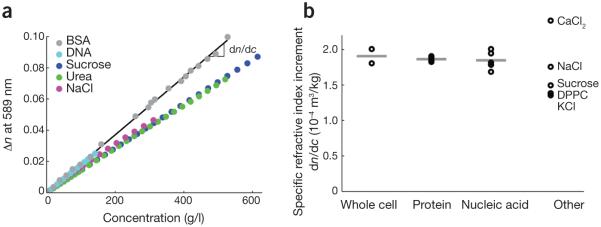
Specific refractive index increment of biomolecules. (a) Changes in refractive index, n, of various biomolecules in solution relative to that of the solvent (water) as a function of biomolecule mass concentration. The slope of this line, dn/dc, is the specific refractive increment used in determinations of cell dry mass from quantitative phase images. (b) Representative specific refractive index increments of selected biomolecule groups, with the averages of these selected values indicated as horizontal bars. Data are shown for whole-cell average values,, proteins (multiprotein average–, bovine serum albumin (BSA) and Evans bacteriological peptone), nucleic acids (DNA– and RNA,) and other components (CaCl2, NaCl, KCl, sucrose and dipalmitoyl phosphatidylcholine (DPPC), a common phospolipid).
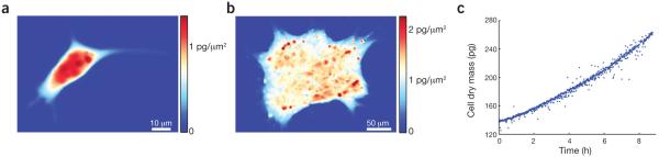
Live-cell mass quantification with interference microscopy. (a,b) Mass distributions within a single, adherent mouse L-cell fibroblast (a) and a multicellular human pluripotent stem cell colony (b) measured with phase-shifting interferometry. The color scale corresponds to mass per unit area, showing the intracellular or intracolony distribution of mass (red, high mass; blue, low mass or background level). (c) Mass distribution images captured by time-lapse interference microscopy can be integrated over the area of a cell or colony to yield mass over time data. A mass-versus-time plot is shown for a single, nonadherent mouse CH12 erythroleukemia cell measured with phase-shifting interferometry.
References
-
- Lloyd AC. The regulation of cell size. Cell. 2013;154:1194–1205. Recent review summarizing the current understanding of cell size control mechanisms. - PubMed
-
- O’Farrell PH. In: Cell Growth: Control of Cell Size. Hall MN, Raff M, Thomas G, editors. Cold Spring Harbor Press; 2004. pp. 1–22. Ch. 1.
-
- Mitchison JM. The growth of single cells: I. Schizosaccharomyces pombe. Exp. Cell Res. 1957;13:244–262. - PubMed
Publication types
MeSH terms
Grants and funding
- R01 CA090571/CA/NCI NIH HHS/United States
- PN2 EY018228/EY/NEI NIH HHS/United States
- K25CA157940/CA/NCI NIH HHS/United States
- R01 CA156674/CA/NCI NIH HHS/United States
- R01CA90571/CA/NCI NIH HHS/United States
- T32 CA009120/CA/NCI NIH HHS/United States
- PN2EY018228/EY/NEI NIH HHS/United States
- P01GM081621/GM/NIGMS NIH HHS/United States
- R01 CA185189/CA/NCI NIH HHS/United States
- R01 CA107300/CA/NCI NIH HHS/United States
- R01 GM073981/GM/NIGMS NIH HHS/United States
- R01CA156674/CA/NCI NIH HHS/United States
- R01CA185189/CA/NCI NIH HHS/United States
- K25 CA157940/CA/NCI NIH HHS/United States
- P01 GM081621/GM/NIGMS NIH HHS/United States
- R01GM073981/GM/NIGMS NIH HHS/United States
LinkOut - more resources
Full Text Sources
Other Literature Sources
