

A Case of the Intrapulmonary Spread of Recurrent Respiratory Papillomatosis With Malignant Transformation
- PMID: 25423295
- PMCID: PMC4495862
- DOI: 10.1097/MAJ.0000000000000370
A Case of the Intrapulmonary Spread of Recurrent Respiratory Papillomatosis With Malignant Transformation
Abstract
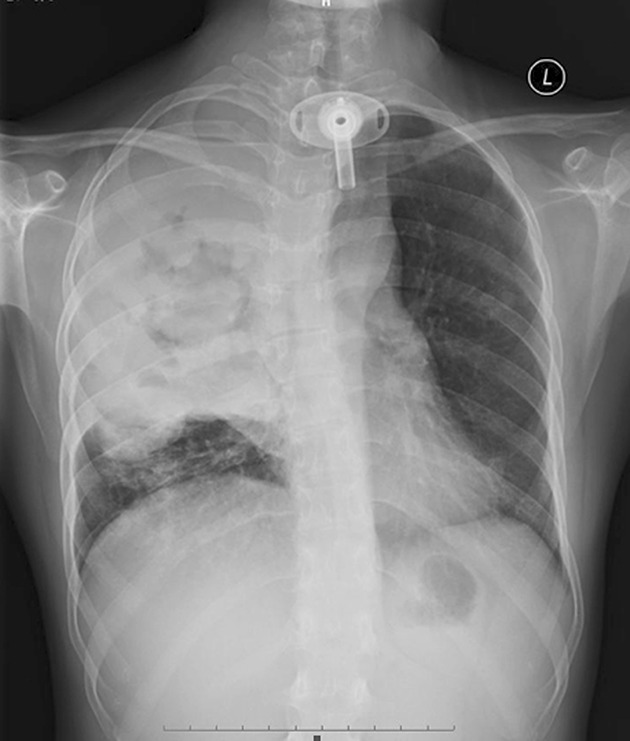
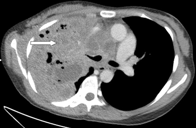
Objectives: To describe an individual with recurrent respiratory papillomatosis that extended into the lung parenchyma and underwent malignant transformation and to discuss the characteristic imaging findings associated with this condition.
Methods: The clinical presentation of an individual with this unusual malignant transformation was reviewed. A literature search was performed to characterize the epidemiology, imaging findings and management of this condition.
Results: The patient underwent 30 courses of surgery over 21 years and presented disseminated pulmonary papilloma after childbirth. The interval between dissemination into the lung and malignant transformation was 2.5 years. The tracheal papilloma was positive for type 6 of human papilloma virus (HPV-6). She died because she refused further treatment.
Conclusions: The clinician should have a high index of suspicion for lung papillomatosis in patients with a tracheotomy. Appropriate diagnostic imaging studies will be helpful in reaching this diagnosis and determining whether a malignancy exists. Treatment options have limited success when lung papillomatosis becomes malignant.
Conflict of interest statement
The authors have no financial or other conflicts of interest to disclose.
Figures
Similar articles
-
Natural History and Malignant Transformation in Recurrent Respiratory Papillomatosis: Human Papillomavirus (HPV), Dysplasia and an Autopsy Review.Fetal Pediatr Pathol. 2015 Apr;34(2):80-90. doi: 10.3109/15513815.2014.968271. Epub 2014 Oct 29. Fetal Pediatr Pathol. 2015. PMID: 25353697 Review.
-
Metastatic squamous-cell carcinoma of the lung arising in a 12-year-old boy with juvenile recurrent respiratory papillomatosis of neonatal onset.Turk Patoloji Derg. 2014;30(2):133-6. doi: 10.5146/tjpath.2014.01238. Turk Patoloji Derg. 2014. PMID: 24638196
-
Squamous cell carcinoma arising in recurrent respiratory papillomatosis with pulmonary involvement: emerging common pattern of clinical features and human papillomavirus serotype association.Mod Pathol. 2000 Aug;13(8):914-8. doi: 10.1038/modpathol.3880164. Mod Pathol. 2000. PMID: 10955460
-
[Two cases of juvenile-onset and adult-onset recurrent respiratory papillomatosis].Nihon Kokyuki Gakkai Zasshi. 2011 Sep;49(9):667-73. Nihon Kokyuki Gakkai Zasshi. 2011. PMID: 22073613 Japanese.
-
Malignant transformation of respiratory papillomatosis in a solid-organ transplant patient: case report and literature review.Ann Otol Rhinol Laryngol. 2013 Jul;122(7):457-60. doi: 10.1177/000348941312200708. Ann Otol Rhinol Laryngol. 2013. PMID: 23951698 Review.
Cited by
-
Recurrent respiratory papillomatosis (RRP) of tracheobronchial tree presenting as lung collapse with malignant transformation after a decade.Respir Med Case Rep. 2023 Aug 1;45:101904. doi: 10.1016/j.rmcr.2023.101904. eCollection 2023. Respir Med Case Rep. 2023. PMID: 37564786 Free PMC article.
-
Investigating cystic lung disease: a respiratory detective approach.Breathe (Sheff). 2020 Jun;16(2):200041. doi: 10.1183/20734735.0041-2020. Breathe (Sheff). 2020. PMID: 33304403 Free PMC article. Review.
-
Laryngotracheobronchial papillomatosis: chest CT findings.J Bras Pneumol. 2017 Jul-Aug;43(4):259-263. doi: 10.1590/S1806-37562016000000351. J Bras Pneumol. 2017. PMID: 29364999 Free PMC article.
-
HPV and Lung Cancer: A Systematic Review.Cancers (Basel). 2024 Sep 28;16(19):3325. doi: 10.3390/cancers16193325. Cancers (Basel). 2024. PMID: 39409943 Free PMC article. Review.
-
Tracheal Papilloma Treated with Cryotherapy and Interferon-α: A Case Report and Review of the Literature.Case Rep Pulmonol. 2015;2015:356796. doi: 10.1155/2015/356796. Epub 2015 Feb 18. Case Rep Pulmonol. 2015. PMID: 25789192 Free PMC article.
References
-
- Donne AJ, Hampson L, Homer JJ, et al. The role of HPV type in recurrent respiratory papillomatosis. Int J Pediatr Otorhinolaryngol 2010;74:7–14. - PubMed
-
- Kramer SS, Wehunt WD, Stocker JT, et al. Pulmonary manifestations of juvenile laryngotracheal papillomatosis. AJR Am J Roentgenol 1985;144:687–94. - PubMed
-
- Dancey DR, Chamberlain DW, Krajden M, et al. Successful treatment of juvenile laryngeal papillomatosis-related multicystic lung disease with cidofovir: case report and review of the literature. Chest 2000;118:1210–4. - PubMed
-
- Cook JR, Hill DA, Humphrey PA, et al. Squamous cell carcinoma arising in recurrent respiratory papillomatosis with pulmonary involvement: emerging common pattern of clinical features and human papillomavirus serotype association. Mod Pathol 2000;13:914–8. - PubMed
-
- Gélinas JF, Manoukian J, Côté A. Lung involvement in juvenile onset recurrent respiratory papillomatosis: a systematic review of the literature. Int J Pediatr Otorhinolaryngol 2008;72:433–52. - PubMed
Publication types
MeSH terms
Supplementary concepts
LinkOut - more resources
Full Text Sources
Medical
