Observational Study
. 2014 Nov-Dec;45(6):496-505.
doi: 10.3928/23258160-20141118-03.

Ultrahigh-speed swept-source OCT angiography in exudative AMD

Observational Study

Ultrahigh-speed swept-source OCT angiography in exudative AMD

Eric Moult et al. Ophthalmic Surg Lasers Imaging Retina. 2014 Nov-Dec.

Abstract

Background and objective: To investigate the potential of ultrahigh-speed swept-source optical coherence tomography angiography (OCTA) to visualize retinal and choroidal vascular changes in patients with exudative age-related macular degeneration (AMD).

Patients and methods: Observational, prospective cross-sectional study. An ultrahigh-speed swept-source prototype was used to perform OCTA of the retinal and choriocapillaris microvasculature in 63 eyes of 32 healthy controls and 19 eyes of 15 patients with exudative AMD.

Main outcome measure: qualitative comparison of the retinal and choriocapillaris microvasculature in the two groups.

Results: Choroidal neovascularization (CNV) was clearly visualized in 16 of the 19 eyes with exudative AMD, located above regions of severe choriocapillaris alteration. In 14 of these eyes, the CNV lesions were surrounded by regions of choriocapillaris alteration.

Conclusion: OCTA may offer noninvasive monitoring of the retinal and choriocapillaris microvasculature in patients with CNV, which may assist in diagnosis and monitoring.


Figures

Figure 1
Figure 1
(A, C) OCT angiograms of a 35-year-old healthy participant. (B, D) OCT angiograms of a 68-year-old healthy participant. (A, B) OCT retinal angiograms over a 6 mm × 6 mm area centered at the fovea. (C, D) OCT choriocapillaris angiograms over the same 6 mm × 6 mm area extracted from the same volumetric scans as used in A and B. Note that the thick retinal vasculature casts angiographic shadows onto the choriocapillaris angiogram. Scale bars: 1 mm.
Figure 2
Figure 2
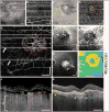
An 87-year-old patient with a choroidal neovascularization (CNV) lesion exhibiting both occult and classic components. (A) En face mean projection of entire OCT intensity volume. (B) En face projection of the OCT angiogram volume through the depths spanned by the retinal vasculature. (C) En face slice of the OCT intensity volume at the depth of the choriocapillaris. The white dashed contour encircles an area of lower intensity. (D) En face slice of the OCT angiogram volume at the depth of the choriocapillaris. (E) En face projection of the OCT angiogram volume through the depths spanned by the CNV. The red outlining corresponds to the occult components of the lesion, while the yellow outlining corresponds to the classic components. The arrows point to motion artifacts that appear as straight white lines through the image. (F) Early-phase FA. Arrows point to the classic component. (G) Early-phase ICGA. Arrows point to the occult component. (H) Late-phase FA. Arrows point to area of stippled hyperfluorescence. (I) OCT retinal thickness map. (J-K) OCT intensity and OCT angiogram cross-sections, respectively, corresponding to the white dashed horizontal lines in (E). The bracket in (J) roughly spans the depths in which the OCT angiogram B-scan is uninterpretable due to shadowing from the choriocapillaris and choroidal vasculature above. The yellow arrows in K indicate the thickness of the choroid away from the lesion, the white arrows demarcate an area of increased light penetration due to RPE atrophy, and the blue arrows correspond to a region of shadowing from the CNV lesion. All OCT images are from a 6 mm × 6 mm area, and all scale bars are 1 mm. All OCT and OCT angiogram images were generated from a single volumetric scan.
Figure 3
Figure 3
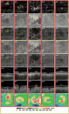
(A-E) Each column corresponds to a different exudative CNV patient. Row 1 displays en face projections of the OCTA volumes through the depths spanned by the CNV. The lesions are indicated by yellow/red outlining. Yellow outlining corresponds to a component that is suspected to be classic; red outlining corresponds to a component that is suspected to be occult. Row 2 shows en face slices of the OCT intensity volume at the depth of the choriocapillaris; row 3 shows the corresponding en face slices of the OCTA volume. Row 4 shows en face slices of the OCT intensity volume at 35 μm beneath the choriocapillaris; row 5 shows the corresponding en face slices of the OCTA volume which shows OCTA shadowing from the choriocapillaris. Rows 6 and 7 show intensity and angiography B-scans, respectively, corresponding to the horizontal and vertical dashed white lines of the first row. The white contour of the row 6 shows the segmentation of Bruch's membrane. Row 8 shows OCT retinal thickness maps. All images were acquired from 6 mm × 6 mm areas. All scale bars are 1 mm.

References

    1. Rosenfeld PJ, Moshfeghi AA, Puliafito CA. Optical coherence tomography findings after an intravitreal injection of bevacizumab (Avastin) for neovascular age-related macular degeneration. Ophthalmic Surg Lasers Imaging Retina. 2005;36(4):331–335. - PubMed
    1. Kaiser PK, Blodi BA, Shapiro H, et al. Angiographic and optical coherence tomographic results of the MARINA study of ranibizumab in neovascular age-related macular degeneration. Ophthalmology. 2007;114(10):1868–1875. - PubMed
    1. Fung AE, Lalwani GA, Rosenfeld PJ, et al. An optical coherence tomography-guided, variable dosing regimen with intravitreal ranibizumab (lucentis) for neovascular age-related macular degeneration. Am J Ophthalmol. 2007;143(4):566–583. - PubMed
    1. Lalwani GA, Rosenfeld PJ, Fung AE, et al. A variable-dosing regimen with intravitreal ranibizumab for neovascular age-related macular degeneration: year 2 of the PrONTO Study. Am J Ophthalmol. 2009;148(1):43–58. - PubMed
    1. Hong YJ, Miura M, Makita S, et al. Noninvasive investigation of deep vascular pathologies of exudative macular diseases by high-penetration optical coherence angiography. Invest Ophthalmol Vis Sci. 2013;54(5):3621–3631. - PubMed

Publication types

MeSH terms