. 2014 Nov 26:4:7196.
doi: 10.1038/srep07196.

The Hippo pathway as a target of the Drosophila DRE/DREF transcriptional regulatory pathway

Affiliations

The Hippo pathway as a target of the Drosophila DRE/DREF transcriptional regulatory pathway

Nicole Vo et al. Sci Rep. .

Abstract

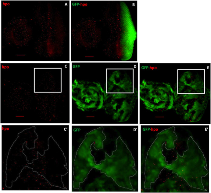
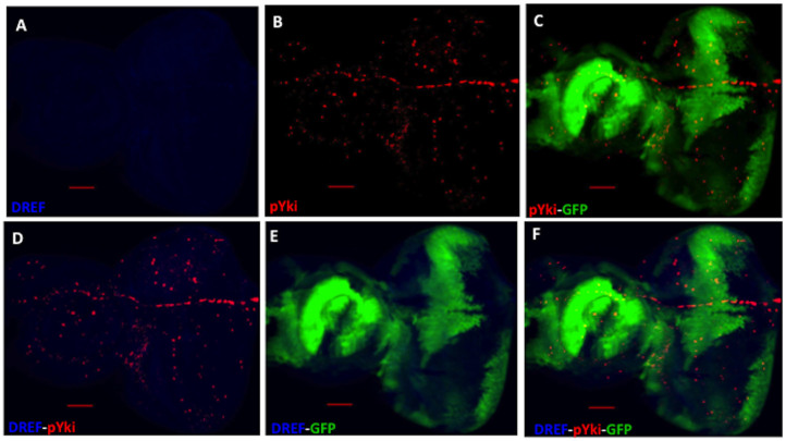
The DRE/DREF transcriptional regulatory system has been demonstrated to activate a wide variety of genes with various functions. In Drosophila, the Hippo pathway is known to suppress cell proliferation by inducing apoptosis and cell cycle arrest through inactivation of Yorkie, a transcription co-activator. In the present study, we found that half dose reduction of the hippo (hpo) gene induces ectopic DNA synthesis in eye discs that is suppressed by overexpression of DREF. Half reduction of the hpo gene dose reduced apoptosis in DREF-overexpressing flies. Consistent with these observations, overexpression of DREF increased the levels of hpo and phosphorylated Yorkie in eye discs. Interestingly, the diap1-lacZ reporter was seen to be significantly decreased by overexpression of DREF. Luciferase reporter assays in cultured S2 cells revealed that one of two DREs identified in the hpo gene promoter region was responsible for promoter activity in S2 cells. Furthermore, endogenous hpo mRNA was reduced in DREF knockdown S2 cells, and chromatin immnunoprecipitation assays with anti-DREF antibodies proved that DREF binds specifically to the hpo gene promoter region containing DREs in vivo. Together, these results indicate that the DRE/DREF pathway is required for transcriptional activation of the hpo gene to positively control Hippo pathways.


Figures

Figure 1
Figure 1. Half reduction of the hpo gene dose enhances cell proliferation which can be suppressed by over-expression of DREF.
(A) Eye imaginal discs of flies overexpressing UAS-GFP are stained with anti-GFP antibody (Red) and DAPI (Blue). (B) Eye discs expressing UAS-DREF are stained with anti-DREF antibody (Red) and DAPI (Blue). (For cell proliferation, eye imaginal discs were labeled with EdU (Red). (C) GMR-GAL4/+; UAS-GFP/+; +. (D) GMR-GAL4/+; UAS-DREF/+; +. (E) w*; +; hpoKC202/CyO. (F) GMR-GAL4/+; UAS-DREF/+; hpoKC202/+. (G) Quantification of the number of ectopic EdU cells in the posterior region of the eye discs. ***p<0.001, ****p<0.0001. White arrowheads indicate morphogenetic furrow (MF). The white border line indicates the posterior region (a) indicates anterior, (p) indicates posterior. Scale bars are 40 μm. The flies were reared at 28°C.
Figure 2
Figure 2. Half dose reduction of the hpo gene reduces the cell death program in DREF over-expression flies.
Cell Event Caspase-3/7 Green Detection Reagent assays were performed in eye imaginal discs (Green). (A) GMR-GAL4/+; UAS-GFP/+; +. (B) GMR-GAL4/+; UAS-DREF/+; +. (C) w*; hpoKC202/CyO; +. (D) GMR-GAL4/+; UAS-DREF/+; hpoKC202/+. (E) Quantification of the number of apoptotic cells in the posterior region of the eye discs. ***p<0.001, ****p<0.0001. White arrowheads indicate the morphogenetic furrow (MF). The white border line indicates the posterior region. (a) indicates anterior, and (p) indicates posterior. Scale bars are 40 μm. The flies were reared at 28°C.
Figure 3
Figure 3. Over-expression of DREF significantly enhances Hpo signals in eye imaginal discs.
Eye imaginal discs were immunostained with anti-hpo antibodies (Red). (A) GMR-GAL4/+; UAS-GFP/+; +. (B) GMR-GAL4/+; UAS-DREF/+; +. (C) w*; +; hpoKC202/CyO. (D) GMR-GAL4/+; UAS-DREF/+; hpoKC202/+. (E) Quantification of the number of Hpo positive signals in the posterior region of the eye discs. ***p<0.001, ****p<0.0001. White arrowheads indicate the morphogenetic furrow (MF). The white border line indicates the posterior region (a) indicates anterior, (p) indicates posterior. Scale bars are 40 μm. The flies were reared at 28°C.
Figure 4
Figure 4. Confirmation of DREF knockdown in flip-out experiments.
(A) Eye imaginal discs of hs-flp; Act5C>FRT y FRT>GAL4, UAS-DREFIR flies are stained with anti-DREF antibody (Blue). (B) Cells expressing DREFdsRNA are marked with GFP (Green). (C) Merged image of anti-DREF and GFP signals in DREF knockdown eye discs. (A'), (B') and (C') are larger images of white box areas in (A), (B), and (C), respectively. In the control flies expressing GAL4 alone, the eye discs were stained with anti-DREF antibody (D) and anti-GFP antibody (E). (F) Merge image of anti-DREF and GFP signals in the control eye discs. The bars indicate 40 μm.
Figure 5
Figure 5. Expression of DREF dsRNA reduces hippo levels in eye discs.
(A) Eye imaginal discs of GMR>GFP flies are stained with anti-hpo antibody (Red). (B) Merge image of anti-hpo and GFP signals (Green) of the flies overexpressing GFP in the posterior region. (C) Eye discs of the flip-out experiment are stained with anti-hpo antibody (Red). (D) Cells expressing DREFdsRNA are marked with GFP (Green). (E) Merged image of anti-hpo and GFP signals in DREF knockdown eye discs. (C'), (D') and (E') are larger images of white box areas in (C), (D), and (E), respectively. Scale bars indicate 40 μm.
Figure 6
Figure 6. Over-expression of DREF increases the phospho-Yki level and reduces diap1-lacZ signals in eye imaginal discs.
Eye imaginal discs were immunostained with anti-phospho-Yki antibodies. (Red) (A) GMR-GAL4/+; UAS-GFP/+; +. (B) GMR-GAL4/+; UAS-DREF/+; +. (C) w*; +;hpoKC202/CyO. (D) GMR-GAL4/+; UAS-DREF/+; hpoKC202/+. (E) Quantification of the number of positive phospho-Yki signals in the posterior region of the eye discs. To examine expression of the diap1-lacZ reporter, eye discs were stained with anti-β-galactosidase antibody (Red) and DAPI (Blue). (F) GMR-GAL4/+; +; diap1-lacZ/+; (G) GMR-GAL4/+; UAS-DREF/+; diap1-lacZ/+. (H) Quantification of the number of β-galactosidase positive signals in the region posterior to the morphogenetic furrow. *p<0.05, ***p<0.001, ****p<0.0001. White arrowheads indicate the morphogenetic furrow (MF). The white border line indicates the posterior region. (a) indicates anterior, and (p) indicates posterior. Scale bars are 40 μm. The flies were reared at 28°C.
Figure 7
Figure 7. Expression of DREF dsRNA reduces phospho-Yki levels in eye discs.
Eye imaginal discs of hs-flp; Act5C>FRT y FRT>GAL4, UAS-DREFIR flies are stained with anti-DREF antibody (Blue) (A) and with anti-phospho Yki antibody (Red) (C). (B) Merged image of DREF and phospho-Yki signals in DREF knockdown eye discs. (D) Cells expressing DREFdsRNA are marked with GFP (Green). (E) Merged image of phospho-Yki and GFP signals. (F) Merged image of DREF, phospho-Yki and GFP signals. (A'–F') larger images of white box areas in (A–F). The white arrows indicate both DREF-positive and phospho Yki-positive cells in GFP clone area. The bars indicate 40 μm.
Figure 8
Figure 8. Distribution of endogenous phospho-Yki in the control flip-out experiments.
Eye imaginal discs of hs-flp; Act5C>FRT y FRT>GAL4, UAS-GFP flies are stained with anti-DREF antibody (Blue) (A) and with anti-phospho Yki antibody (Red) (B). (C) Merged image of phospho-Yki and GFP signals. (D) Merged image of DREF and phospho-Yki signals. (E) Merged image of DREF and GFP signals. (F) Merged image of DREF, phospho-Yki and GFP signals. The endogenous phospho-Yki signals scattered throughout the whole eye discs and there was no change in the level of DREF signals. The bars indicate 40 μm.
Figure 9
Figure 9. The hpo gene carries DRE and DRE-like sequences in the 5′-flanking region.
(A) Schematic of the 5′-flanking region of the hpo gene. The transcription initiation site is indicated by the arrow and designated as +1. The positions and nucleotide sequences of DRE1 and DRE2 are shown. Arrowheads indicate positions of the primers used for real-time PCR reactions. (B) Crosslinked chromatin of S2 cells was immunoprecipitated with either anti-DREF IgG or control rabbit IgG. Genomic regions containing hpoDRE1, hpoDRE2, and wtsDRE1, 2 were amplified by PCR, and compared with amplicons from immunoprecipitates with control rabbit IgG.
Figure 10
Figure 10. DREF plays essential roles in hpo gene promoter activity in cultured S2 cells.
(A) Schematic features of the hpo promoter-luciferase fusion plasmid hpo-WT-luc and its base-substituted derivatives (hpo-DRE1mut-luc, hpo-DRE2mut-luc, hpo-DREallmut-luc). DRE is represented by an open box and mutated DRE by a closed box. Plasmids were transfected into S2 cells and luciferase activities measured at 48 hours thereafter. Luciferase activity was normalized to Renilla luciferease activity and expressed relative to that of hpo-WT-luc. Mean activities with standard deviations from three independent transfections are shown. (B) Effects of DREFdsRNA treatment on hpo gene promoter activity in S2 cells. Mean activities with standard deviations from three independent transfections are shown. (C) DREFdsRNA treatment reduces endogenous hpo mRNA levels in S2 cells. DREF mRNA, wts mRNA and hpo mRNA in DREF dsRNA-treated cells were measured by quantitative RT-PCR and compared with the values for non-dsRNA treated cells (Mock). mRNA for β-tubulin was used as a negative control and mRNA for wts as a positive control. * p<0.05.
Figure 11
Figure 11. A model of the role of DREF in regulation of Hippo, JNK and p53 pathways inducing apoptosis.
DREF binds to DRE in the hpo promoter region which activates hpo gene transcription. DREF upregulates both JNK and p53 pathways to induce apoptosis. Activation of the Hippo pathway by DREF inactivates Yorkie to further enhance apoptosis.

Similar articles

Cited by

References

    1. Hirose F., Yamaguchi M., Handa H., Inomata Y. & Matsukage A. Novel 8-base pair sequence (Drosophila DNA replication-related element) and specific binding factor involved in the expression of Drosophila genes for DNA polymerase alpha and proliferating cell nuclear antigen. J. Biol. Chem. 268, 2092–2099 (1993). - PubMed
    1. Ohler U., Liao G. C., Niemann H. & Rubin G. M. Computational analysis of core promoters in the Drosophila genome. Genome Biol. 3, RESEARCH0087 (2002). - PMC - PubMed
    1. Matsukage A., Hirose F., Yoo M. A. & Yamaguchi M. The DRE/DREF transcriptional regulatory system: a master key for cell proliferation. Biochim. Biophys. Acta. 1779, 81–89, 10.1016/j.bbagrm.2007.11.011 (2008). - PubMed
    1. Yamaguchi M., Hayashi Y. & Matsukage A. Essential role of E2F recognition sites in regulation of the proliferating cell nuclear antigen gene promoter during Drosophila development. J. Biol. Chem. 270, 25159–25165 (1995). - PubMed
    1. Okudaira K. et al. Transcriptional regulation of the Drosophila orc2 gene by the DREF pathway. Biochim. Biophys. Acta. 1732, 23–30, 10.1016/j.bbaexp.2005.10.009 (2005). - PubMed

Publication types

MeSH terms

Substances

LinkOut - more resources